
Page 56 - Read Online
P. 56
Page 4 of 17 Cevallos et al. Plast Aesthet Res 2023;10:30 https://dx.doi.org/10.20517/2347-9264.2023.01
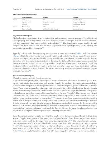
Table 1. Clinical examination findings
Characteristics Arterial Venous
Visual Color Pale Cyanotic, dusky
Capillary Refill Delayed (> 3 secs) Shortened (< 2 secs)
Pinprick Decreased Dark venous outflow
Tactile Temperature Cold Warmth
Tension/Swelling Mild swelling (late finding) Increased turgor
Adjunctive techniques
Medical devices manufacture is an evolving field and an area of ongoing research. The objective of
developing flap monitoring devices is to create compact, portable techniques that can provide consistent,
real-time quantitative data on flap viability and perfusion [43-47] . The data obtained should be objective and
not provider-dependent [48,49] . This data can assist surgeons in assessing flow patterns, quality, severity, and
determining the need for reoperation .
[50]
Typically, techniques for flap monitoring are categorized as either non-invasive [Tables 1 and 2] or invasive
[Table 3]. Multiple devices can be used in combination as adjuncts to the clinical exam. Generally, non-
invasive techniques are more user-friendly, readily available, and cost-effective. Quantitative results that can
be tracked over time enhance the sensitivity of detecting flap failure. Monitoring devices may enable flap
monitoring without direct contact with providers, which was advantageous during the COVID-19
pandemic . However, it is important to note that absolute values may have limitations and lack
[51]
consistency between patients. Further, the use of monitoring machines may entail costs and require
specialized expertise .
[48]
Non-invasive techniques
Photoelectric assessment with Doppler monitoring
Doppler ultrasonography is widely recognized as one of the most effective and commonly used non-
invasive methods for flap monitoring, with acoustic Doppler devices being the most predominant choice.
Acoustic Dopplers operate by emitting sound waves of a specific frequency, phase, and amplitude into the
tissue. These sound waves reflect off moving matter, primarily the red blood cells within the intravascular
arterial and venous system in flaps. The movement of these cells leads to a slight shift in the frequency of the
reflected sound waves, known as the Doppler effect, hence the term “Doppler”. The machine converts and
transmits these reflections into an audible noise, which exhibits a pulsatile pattern in arterial flow and a
quieter, more consistent pattern in venous flow [52-54] . Marking arterial and venous acoustic signals at the
conclusion of the procedure with a single stitch can facilitate post-operative monitoring . Acoustic
[55]
Doppler sonography is a user-friendly technique that requires minimal training, and the devices are widely
available, cost-effective, and highly portable . However, it is important to note that the absence of a signal
[54]
does not always indicate inadequate blood supply for flap viability, and the signal from key perforators may
[53]
be difficult to distinguish from background noise caused by other vessels .
Laser flowmetry is another Doppler-based method employed for flap monitoring, although it differs from
acoustic Doppler by measuring in light waves instead of sound waves . Laser flowmetry probes are secured
[56]
to the skin surface overlying the target vessel using dressings or sutures [57,58] . The data output provides real-
time measurements of skin surface blood velocity and blood flow, which are accurate up to a depth of
8 mm . However, these values only serve as relative indicators of blood flow, and while trends within the
[13]
same patient are reliable, the data can be significantly influenced by various patient- and device-related
factors [57,59] . As a result, there is no clear “cut-off” value suggestive of vascular compromise. Instead,