
Page 30 - Read Online
P. 30
Van Hove et al. Plast Aesthet Res 2023;10:8 https://dx.doi.org/10.20517/2347-9264.2022.59 Page 7 of 14
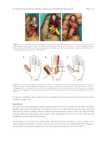
Figure 6. A: This patient was referred 48 hours after replantation for impending venous failure. B: A 1.0 cm × 2.5 cm first dorsal
metacarpal flap was elevated from the dorsum of the index permitting primary closure of the donor site. The flap eased the circular soft
tissue constriction of the thumb and (most importantly) carried two veins restoring the outflow of the thumb (blue arrows). C:
Complete survival at two weeks (© Dr. Piñal 2016) [30] .
Figure 7. A: An extended island flap is designed on the ulnar side of the long finger with an identical defect pattern over the palmar
aspect of the thumb. B: Mobilization of the island flap on its neurovascular pedicle and preparation of the recipient bed on the thumb. C:
Completed transfer of the neurovascular island flap with a full-thickness skin graft on the donor area of the long finger. Care must be
taken to avoid a longitudinal scar along the palmar aspect of the donor finger. Note the area of undermining in the palm (© Dr. Piñal
[6]
2020) .
In summary, this flap is used in patients with considerable bone exposure and who do not wish for more
complex reconstruction.
Nail defects
Not only is the nail an important cosmetic element, but it also allows us to pick up small objects and offers
stability to the pulp when pinching. The problem of nail loss is compounded because it is often associated
with bone loss. Hence, a length problem is added to the functional and cosmetic issues. The nail grows short
and lacks adherence if the entire sterile matrix is lost. The patient may wish to have their nail removed
completely to prevent this from happening.
An alternative is to transfer the sterile matrix and the bone from the hallux as a free onycho-osteo-
cutaneous flap. Remarkably, the matrix of the toe will “meld” with the remaining thumbnail [36,37] [Figure 8].
Microvascular reconstruction appears to be the only method that procures consistent results [38-42] .