
Page 33 - Read Online
P. 33
Page 10 of 14 Van Hove et al. Plast Aesthet Res 2023;10:8 https://dx.doi.org/10.20517/2347-9264.2022.59
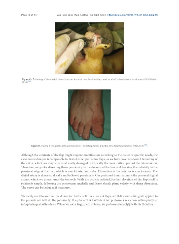
Figure 10. Trimming of the medial side of the toe. Asterisk: medial-sided flap used as a V-Y advancement for closure (©Dr Piñal in
2014) [36] .
Figure 11. Placing a skin graft on the periosteum of the distal phalanx provides for a facsimile nail(©Dr Piñal 2014) [36] .
Although the contents of the flap might require modification according to the patient’s specific needs, the
elevation technique is comparable to that of other partial toe flaps, as we have covered above. Harvesting of
the veins, which are very small and easily damaged, is typically the most critical part of the intervention.
Therefore, we prefer dissecting them proximally in the dorsum of the foot and tracking them distally to the
proximal edge of the flap, which is much faster and safer. Dissection of the arteries is much easier. The
digital artery is dissected distally and followed proximally. Our preferred donor artery is the peroneal digital
artery, which we dissect until the toe web. With the pedicle isolated, further elevation of the flap itself is
relatively simple, following the periosteum medially and flexor sheath plane volarly with sharp dissection.
The nerve can be included if necessary.
We rarely need to sacrifice the donor toe. In the soft tissue variant flaps, a full-thickness skin graft applied to
the periosteum will do the job nicely. If a phalanx is harvested, we perform a resection arthroplasty or
interphalangeal arthrodesis. When we use a large piece of bone, we perform syndactyly with the third toe.