
Page 29 - Read Online
P. 29
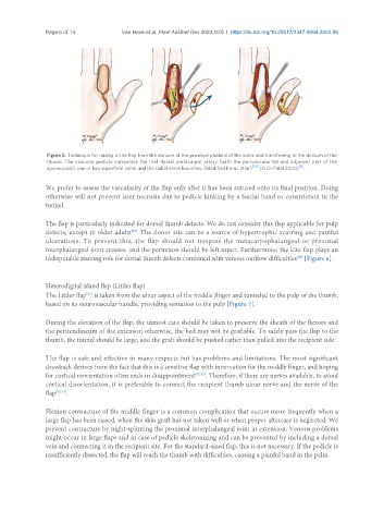
Page 6 of 14            Van Hove et al. Plast Aesthet Res 2023;10:8  https://dx.doi.org/10.20517/2347-9264.2022.59


                Figure 5. Technique for raising a kite flap from the dorsum of the proximal phalanx of the index and transferring to the dorsum of the
                thumb. The vascular pedicle comprises the first dorsal metacarpal artery (with the perivascular fat and adjacent part of the
                                                                                              [6]
                aponeurosis), one or two superficial veins, and the radial nerve branches. (Modified from Littler) [28]  (© Dr Piñal 2020) .

               We prefer to assess the vascularity of the flap only after it has been sutured onto its final position. Doing
               otherwise will not prevent later necrosis due to pedicle kinking by a fascial band or constriction in the
               tunnel.

               The flap is particularly indicated for dorsal thumb defects. We do not consider this flap applicable for pulp
               defects, except in older adults . The donor site can be a source of hypertrophic scarring and painful
                                          [29]
               ulcerations. To prevent this, the flap should not trespass the metacarpophalangeal or proximal
               interphalangeal joint creases, and the peritenon should be left intact. Furthermore, the kite flap plays an
                                                                                             [30]
               indisputable starring role for dorsal thumb defects combined with venous outflow difficulties  [Figure 6].


               Heterodigital island flap (Littler flap)
                            [31]
               The Littler flap  is taken from the ulnar aspect of the middle finger and tunneled to the pulp of the thumb,
               based on its neurovascular bundle, providing sensation to the pulp [Figure 7].

               During the elevation of the flap, the utmost care should be taken to preserve the sheath of the flexors and
               the peritendineum of the extensor; otherwise, the bed may not be graftable. To safely pass the flap to the
               thumb, the tunnel should be large, and the graft should be pushed rather than pulled into the recipient side.

               The flap is safe and effective in many respects but has problems and limitations. The most significant
               drawback derives from the fact that this is a sensitive flap with innervation for the middle finger, and hoping
               for cortical reorientation often ends in disappointment [32-35] . Therefore, if there are nerves available, to avoid
               cortical disorientation, it is preferable to connect the recipient thumb ulnar nerve and the nerve of the
               flap [32,33] .


               Flexion contracture of the middle finger is a common complication that occurs more frequently when a
               large flap has been raised, when the skin graft has not taken well or when proper aftercare is neglected. We
               prevent contracture by night-splinting the proximal interphalangeal joint in extension. Venous problems
               might occur in large flaps and in case of pedicle skeletonizing and can be prevented by including a dorsal
               vein and connecting it in the recipient site. For the standard-sized flap, this is not necessary. If the pedicle is
               insufficiently dissected, the flap will reach the thumb with difficulties, causing a painful band in the palm.