
Page 70 - Read Online
P. 70
Page 66 Tang et al. Neuroimmunol Neuroinflammation 2021;8:64-9 I http://dx.doi.org/10.20517/2347-8659.2020.28
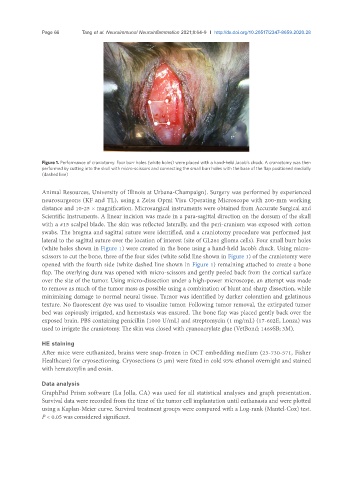
Figure 1. Performance of craniotomy. Four burr holes (white holes) were placed with a hand-held Jacob’s chuck. A craniotomy was then
performed by cutting into the skull with micro-scissors and connecting the small burr holes with the base of the flap positioned medially
(dashed line)
Animal Resources, University of Illinois at Urbana-Champaign). Surgery was performed by experienced
neurosurgeons (KF and TL), using a Zeiss Opmi Visu Operating Microscope with 200-mm working
distance and 10-25 × magnification. Microsurgical instruments were obtained from Accurate Surgical and
Scientific Instruments. A linear incision was made in a para-sagittal direction on the dorsum of the skull
with a #15 scalpel blade. The skin was reflected laterally, and the peri-cranium was exposed with cotton
swabs. The bregma and sagittal suture were identified, and a craniotomy procedure was performed just
lateral to the sagittal suture over the location of interest (site of GL261 glioma cells). Four small burr holes
(white holes shown in Figure 1) were created in the bone using a hand-held Jacob’s chuck. Using micro-
scissors to cut the bone, three of the four sides (white solid line shown in Figure 1) of the craniotomy were
opened with the fourth side (white dashed line shown in Figure 1) remaining attached to create a bone
flap. The overlying dura was opened with micro-scissors and gently peeled back from the cortical surface
over the site of the tumor. Using micro-dissection under a high-power microscope, an attempt was made
to remove as much of the tumor mass as possible using a combination of blunt and sharp dissection, while
minimizing damage to normal neural tissue. Tumor was identified by darker coloration and gelatinous
texture. No fluorescent dye was used to visualize tumor. Following tumor removal, the extirpated tumor
bed was copiously irrigated, and hemostasis was ensured. The bone flap was placed gently back over the
exposed brain. PBS containing penicillin (1000 U/mL) and streptomycin (1 mg/mL) (17-602E, Lonza) was
used to irrigate the craniotomy. The skin was closed with cyanoacrylate glue (VetBond; 1469SB; 3M).
HE staining
After mice were euthanized, brains were snap-frozen in OCT embedding medium (23-730-571, Fisher
Healthcare) for cryosectioning. Cryosections (5 µm) were fixed in cold 95% ethanol overnight and stained
with hematoxylin and eosin.
Data analysis
GraphPad Prism software (La Jolla, CA) was used for all statistical analyses and graph presentation.
Survival data were recorded from the time of the tumor cell implantation until euthanasia and were plotted
using a Kaplan-Meier curve. Survival treatment groups were compared with a Log-rank (Mantel-Cox) test.
P < 0.05 was considered significant.