
Page 87 - Read Online
P. 87
Yoshimura et al. Neuroimmunol Neuroinflammation 2020;7:264-76 I http://dx.doi.org/10.20517/2347-8659.2020.22 Page 265
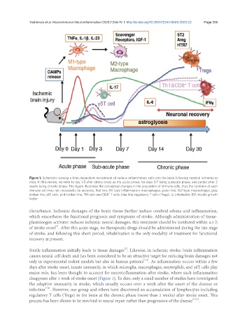
Figure 1. Schematic viewing a time-dependent recruitment of various inflammatory cells into the brain following cerebral ischemia in
mice. In this review, we refer to day 1-3 after stroke onset as the acute phase, for days 3-7 being subacute phase, and period after 2
weeks being chronic phase. This figure illustrates the conceptual changes in the population of immune cells, thus the numbers of each
immune cell may not necessarily be accurate. Red line; M1 type inflammatory macrophages, green line; M2 type macrophages, gray
+
broken line; γδT cells, pink broken line; Th1 cells and CD8 T cells, blue line; regulatory T cells (Tregs). IL: interleukin; IGF: insulin growth
factor
disturbance. Ischemic damages of the brain tissue further induce cerebral edema and inflammation,
which exacerbates the functional prognosis and symptoms of stroke. Although administration of tissue-
plasminogen activator reduces ischemic neural damages, this treatment should be instituted within 4.5 h
[2]
of stroke onset . After this acute stage, no therapeutic drugs should be administered during the late stage
of stroke, and following this short period, rehabilitation is the only modality of treatment for functional
recovery at present.
[3]
Sterile inflammation initially leads to tissue damages . Likewise, in ischemic stroke, brain inflammation
causes neural cell death and has been considered to be an attractive target for reducing brain damages not
[4-6]
only in experimental rodent models but also in human patients . As inflammation occurs within a few
days after stroke onset, innate immunity, in which microglia, macrophages, neutrophils, and γδT cells play
major role, has been thought to account for neuroinflammation after stroke, where such inflammation
disappears after 1 week of stroke onset [Figure 1]. To date, only a small number of studies have investigated
the adaptive immunity in stroke, which usually occurs over a week after the onset of the disease or
infection . However, our group and others have discovered an accumulation of lymphocytes including
[7,8]
regulatory T cells (Tregs) in the brain at the chronic phase (more than 2 weeks) after stroke onset. This
process has been shown to be involved in neural repair rather than progression of the disease [9-13] .