
Page 31 - Read Online
P. 31
Walker. Neuroimmunol Neuroinflammation 2020;7:194-214 I http://dx.doi.org/10.20517/2347-8659.2020.09 Page 209
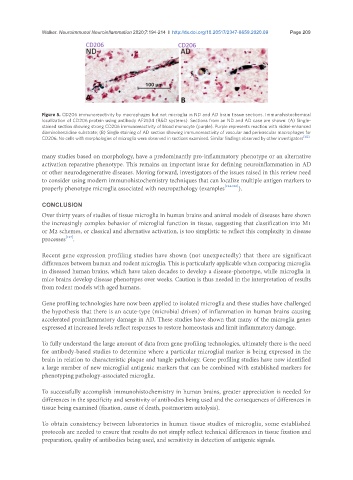
Figure 5. CD206 immunoreactivity by macrophages but not microglia in ND and AD brain tissue sections. Immunohistochemical
localization of CD206 protein using antibody AF2534 (R&D systems). Sections from an ND and AD case are shown: (A) Single-
stained section showing strong CD206 immunoreactivity of blood monocyte (purple). Purple represents reaction with nickel-enhanced
diaminobenzidine substrate; (B) Single staining of AD section showing immunoreactivity of vascular and perivascular macrophages for
CD206. No cells with morphologies of microglia were observed in sections examined. Similar findings observed by other investigators [122]
many studies based on morphology, have a predominantly pro-inflammatory phenotype or an alternative
activation reparative phenotype. This remains an important issue for defining neuroinflammation in AD
or other neurodegenerative diseases. Moving forward, investigators of the issues raised in this review need
to consider using modern immunohistochemistry techniques that can localize multiple antigen markers to
properly phenotype microglia associated with neuropathology (examples [124-126] ).
CONCLUSION
Over thirty years of studies of tissue microglia in human brains and animal models of diseases have shown
the increasingly complex behavior of microglial function in tissue, suggesting that classification into M1
or M2 schemes, or classical and alternative activation, is too simplistic to reflect this complexity in disease
processes [127] .
Recent gene expression profiling studies have shown (not unexpectedly) that there are significant
differences between human and rodent microglia. This is particularly applicable when comparing microglia
in diseased human brains, which have taken decades to develop a disease-phenotype, while microglia in
mice brains develop disease phenotypes over weeks. Caution is thus needed in the interpretation of results
from rodent models with aged humans.
Gene profiling technologies have now been applied to isolated microglia and these studies have challenged
the hypothesis that there is an acute-type (microbial driven) of inflammation in human brains causing
accelerated proinflammatory damage in AD. These studies have shown that many of the microglia genes
expressed at increased levels reflect responses to restore homeostasis and limit inflammatory damage.
To fully understand the large amount of data from gene profiling technologies, ultimately there is the need
for antibody-based studies to determine where a particular microglial marker is being expressed in the
brain in relation to characteristic plaque and tangle pathology. Gene profiling studies have now identified
a large number of new microglial antigenic markers that can be combined with established markers for
phenotyping pathology-associated microglia.
To successfully accomplish immunohistochemistry in human brains, greater appreciation is needed for
differences in the specificity and sensitivity of antibodies being used and the consequences of differences in
tissue being examined (fixation, cause of death, postmortem autolysis).
To obtain consistency between laboratories in human tissue studies of microglia, some established
protocols are needed to ensure that results do not simply reflect technical differences in tissue fixation and
preparation, quality of antibodies being used, and sensitivity in detection of antigenic signals.