
Page 29 - Read Online
P. 29
Walker. Neuroimmunol Neuroinflammation 2020;7:194-214 I http://dx.doi.org/10.20517/2347-8659.2020.09 Page 207
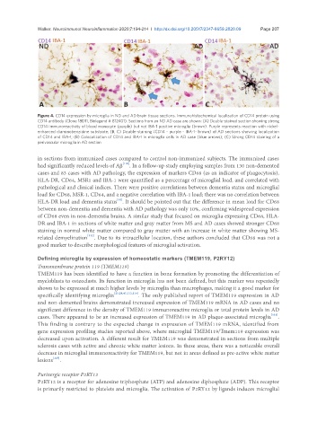
Figure 4. CD14 expression by microglia in ND and AD brain tissue sections. Immunohistochemical localization of CD14 protein using
CD14 antibody (Clone 18D11, Biolegend # 812401). Sections from an ND AD case are shown: (A) Double-stained section showing strong
CD14 immunoreactivity of blood monocyte (purple) but not IBA-1 positive microglia (brown). Purple represents reaction with nickel-
enhanced diaminobenzidine substrate; (B, C) Double-staining (CD14 - purple - IBA-1- brown) of AD sections showing localization
of CD14 and IBA-1; (B) Colocalization of CD14 and IBA-1 in microglia cells in AD case (blue arrows); (C) Strong CD14 staining of a
perivascular microglia in AD section
in sections from immunized cases compared to control non-immunized subjects. The immunized cases
had significantly reduced levels of Aβ [110] . In a follow-up study employing samples from 130 non-demented
cases and 83 cases with AD pathology, the expression of markers CD68 (as an indicator of phagocytosis),
HLA-DR, CD64, MSR1 and IBA-1 were quantified as a percentage of microglial load, and correlated with
pathological and clinical indices. There were positive correlations between dementia status and microglial
load for CD68, MSR-1, CD64, and a negative correlation with IBA-1 load; there was no correlation between
[34]
HLA-DR load and dementia status . It should be pointed out that the difference in mean load for CD68
between non-dementia and dementia with AD pathology was only 10%, confirming widespread expression
of CD68 even in non-dementia brains. A similar study that focused on microglia expressing CD68, HLA-
DR and IBA-1 in sections of white matter and gray matter from MS and AD cases showed stronger CD68
staining in normal white matter compared to gray matter with an increase in white matter showing MS-
related demyelination [112] . Due to its intracellular location, these authors concluded that CD68 was not a
good marker to describe morphological features of microglial activation.
Defining microglia by expression of homeostatic markers (TMEM119, P2RY12)
Transmembrane protein 119 (TMEM119)
TMEM119 has been identified to have a function in bone formation by promoting the differentiation of
myeloblasts to osteoclasts. Its function in microglia has not been defined, but this marker was repeatedly
shown to be expressed at much higher levels by microglia than macrophages, making it a good marker for
specifically identifying microglia [25,28,47,113,114] . The only published report of TMEM119 expression in AD
and non-demented brains demonstrated increased expression of TMEM119 mRNA in AD cases and no
significant difference in the density of TMEM119 immunoreactive microglia or total protein levels in AD
cases. There appeared to be an increased expression of TMEM119 in AD plaque-associated microglia [113] .
This finding is contrary to the expected change in expression of TMEM119 mRNA, identified from
gene expression profiling studies reported above, where microglial TMEM119/Tmem119 expression was
decreased upon activation. A different result for TMEM119 was demonstrated in sections from multiple
sclerosis cases with active and chronic white matter lesions. In these areas, there was a noticeable overall
decrease in microglial immunoreactivity for TMEM119, but not in areas defined as pre-active white matter
lesions [115] .
Purinergic receptor P2RY12
P2RY12 is a receptor for adenosine triphosphate (ATP) and adenosine diphosphate (ADP). This receptor
is primarily restricted to platelets and microglia. The activation of P2RY12 by ligands induces microglial