
Page 25 - Read Online
P. 25
Walker. Neuroimmunol Neuroinflammation 2020;7:194-214 I http://dx.doi.org/10.20517/2347-8659.2020.09 Page 203
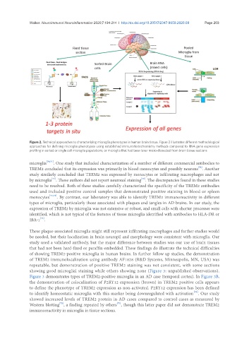
Figure 2. Technical approaches to characterizing microglia phenotypes in human brain tissue. Figure 2 illustrates different methodological
approaches for defining microglia phenotypes using established immunohistochemistry methods compared to RNA gene expression
profiling in sorted or single cell microglia populations, or microglia that had been laser micro-dissected from brain tissue sections
microglia [70,71] . One study that included characterization of a number of different commercial antibodies to
[70]
TREM2 concluded that its expression was primarily in blood monocytes and possibly neurons . Another
study similarly concluded that TREM2 was expressed by monocytes or infiltrating macrophages and not
[71]
[71]
by microglia . These authors did not report neuronal staining . The discrepancies found in these studies
need to be resolved. Both of these studies carefully characterized the specificity of the TREM2 antibodies
used and included positive control samples that demonstrated positive staining in blood or spleen
monocytes [70,71] . By contrast, our laboratory was able to identify TREM2 immunoreactivity in different
types of microglia, particularly those associated with plaques and tangles in AD brains. In our study, the
expression of TREM2 by microglia was not extensive or robust, and small cells with shorter processes were
identified, which is not typical of the features of tissue microglia identified with antibodies to HLA-DR or
IBA-1 .
[72]
These plaque-associated microglia might still represent infiltrating macrophages and further studies would
be needed, but their localization in brain neuropil and morphology were consistent with microglia. Our
study used a validated antibody, but the major difference between studies was our use of brain tissues
that had not been hard-fixed or paraffin-embedded. These findings do illustrate the technical difficulties
of showing TREM2-positive microglia in human brains. In further follow up studies, the demonstration
of TREM2 immunolocalization using antibody AF1828 (R&D Systems, Minneapolis, MN, USA) was
repeatable, but demonstration of positive TREM2 staining was not consistent, with some sections
showing good microglial staining while others showing none (Figure 3: unpublished observations).
Figure 3 demonstrates types of TREM2-positive microglia in an AD case (temporal cortex). In Figure 3B,
the demonstration of colocalization of P2RY12 expression (brown) in TREM2 positive cells appears
to define the phenotype of TREM2 expression as non-activated. P2RY12 expression has been defined
[28]
to identify homeostatic microglia with this marker being downregulated with activation . Our study
showed increased levels of TREM2 protein in AD cases compared to control cases as measured by
[73]
[72]
Western blotting , a finding repeated by others , though this latter paper did not demonstrate TREM2
immunoreactivity in microglia in tissue sections.