
Page 423 - Read Online
P. 423
Davis et al. Neuroimmunol Neuroinflammation 2018;5:50 I http://dx.doi.org/10.20517/2347-8659.2018.60 Page 7 of 11
Unstim. IL-1β Unstim. IL-1β
2.5 1.5
b P = 0.37
IL-1β
US 2.0
p-p65 65 kDa 1.0
p65 65 kDa p-p65/p65 (relative density) 1.5 ab p65/β-tubulin (relative density)
β-tubulin 55 kDa 1.0 a 0.5
10 30 60
Minutes 0.5 a
0.0 0.0
10 30 60 10 30 60
Minutes Minutes
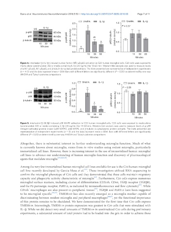
Figure 4. Interleukin-1β (IL-1β)-induced nuclear factor (NF)-κB p65 activation in C20 human microglial cells. C20 cells were exposed to
media alone (unstimulated; US) or media containing IL-1β (20 ng/mL) for 10-60 min. Western blot analysis was used to measure levels
of p-NF-κB p65, NF-κB p65, and β-tubulin in nuclear protein extracts. The blots presented are representative of independent experiments
(n = 4-5) and the data represent mean ± SEM. Bars with different letters are significantly different (P < 0.05) as determined by one-way
ANOVA and Tukey’s pairwise comparisons
Unstim. IL-1β
1.0 1.5 Unstim. IL-1β
IL-1β b P = 0.58
US
p-p38 43 kDa 1.0
0.6
p38 43 kDa p-p38/p38 (relative density) 0.8
β-tubulin 55 kDa 0.4 p38/β-tubulin (relative density)
10 30 60 c 0.5
Minutes 0.2 ac
a
0.0 0.0
10 30 60 10 30 60
Minutes Minutes
Figure 5. Interleukin-1β (IL-1β)-induced p38 MAPK activation in C20 human microglial cells. C20 cells were exposed to media alone
(unstimulated; US) or media containing IL-1β (20 ng/mL) for 10-60 min. Western blot analysis was used to measure levels of p-p38
mitogen-activated protein kinase (p38 MAPK), p38 MAPK, and β-tubulin in cytoplasmic protein extracts. The blots presented are
representative of independent experiments (n = 5) and the data represent mean ± SEM. Bars with different letters are significantly
different (P < 0.05) as determined by one-way ANOVA and Tukey’s pairwise comparisons
Altogether, there is substantial interest in further understanding microglia function. Much of what
is currently known about microglia, stems from in vitro studies using rodent microglia, particularly
immortalized cell lines. However, there is increasing interest in the use of immortalized human microglial
cell lines to advance our understanding of human microglia function and discovery of pharmacological
agents that modulate microglia [37,38,53,54] .
Among the very few immortalized human microglial cell lines available for use is the C20 human microglial
[37]
cell line recently developed by Garcia-Mesa et al. . These investigators utilized RNA sequencing to
confirm the microglial phenotype of C20 cells and they demonstrated that these cells maintain migratory
[37]
capacity and phagocytic activity characteristic of microglia . Furthermore, C20 cells express numerous
microglial surface markers, including cluster of differentiation (CD)11b, CD68, TGFβ receptor (TGFβR),
[37]
and the P2 purinergic receptor, P2RY12, as indicated by immunofluorescence and flow cytometry . While
[55]
+
CD11b macrophages are also present in peripheral tissues , TGFβR and P2RY12 have been suggested
to be microglial specific [56,57] . TMEM119 has also recently emerged as a microglia marker capable of
discriminating between resident microglia and peripheral macrophages [46,47] , yet the functional importance
of this protein remains to be elucidated. We have demonstrated for the first time that C20 cells express
TMEM119. Interestingly, TMEM119 protein expression was greatest in C20 cells that were stimulated with
IL-1β. While we did detect very small amounts of TMEM119 in unstimulated C20 cells during preliminary
experiments, a substantial amount of total protein had to be loaded into the gels in order to achieve these