
Page 422 - Read Online
P. 422
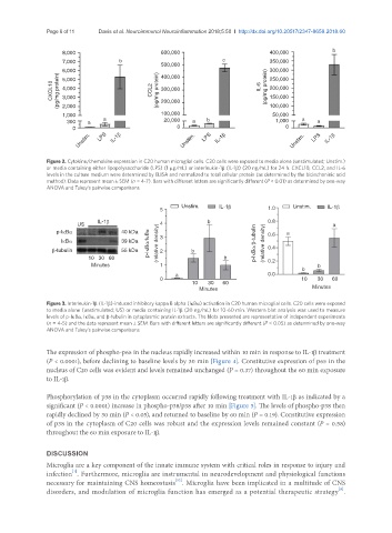
Page 6 of 11                 Davis et al. Neuroimmunol Neuroinflammation 2018;5:50  I  http://dx.doi.org/10.20517/2347-8659.2018.60


                    8,000                         600,000                          400,000            b
                    7,000            b                               c             350,000
                                                  500,000                          300,000
                   (pg/mg protein) 6,000          400,000                       IL-6  (pg/mg protein)  250,000
                    5,000
                 CXCL10  3,000                 CCL2  (pg/mg protein)  200,000      150,000
                    4,000
                                                                                   200,000
                                                  300,000
                    2,000
                                                                                    50,000
                    1,000                         100,000                          100,000
                     300    a   a                  20,000  a    b                    1,000   a   a
                       0                               0                                0
                         Unstim.  LPS  IL-1β            Unstim.  LPS  IL-1β               Unstim.  LPS  IL-1β


               Figure 2. Cytokine/chemokine expression in C20 human microglial cells. C20 cells were exposed to media alone (unstimulated; Unstim.)
               or media containing either lipopolysaccharide (LPS) (1 µg/mL) or interleukin-1β (IL-1β) (20 ng/mL) for 24 h. CXCL10, CCL2, and IL-6
               levels in the culture medium were determined by ELISA and normalized to total cellular protein (as determined by the bicinchoninic acid
               method). Data represent mean ± SEM (n = 4-7). Bars with different letters are significantly different (P < 0.01) as determined by one-way
               ANOVA and Tukey’s pairwise comparisons

                                                        Unstim.     IL-1β                 Unstim.   IL-1β
                                                  5                               1.0
                               IL-1β                            b
                        US                                                                            a
                  p-IκBα              40 kDa                                      0.6   a
                                                  3
                    IκBα              39 kDa  p-IκBα/IκBα  (relative density) 4  p-IκBα/β-tubulin  (relative density) 0.8
                 β-tubulin            55 kDa      2        b                      0.4
                            10   30   60                             b            0.2
                             Minutes              1                                              b
                                                                                             b
                                                      a                           0.0
                                                  0                                          10       30      60
                                                           10       30      60
                                                              Minutes                           Minutes
               Figure 3. Interleukin-1β (IL-1β)-induced inhibitory kappa B alpha (IκBα) activation in C20 human microglial cells. C20 cells were exposed
               to media alone (unstimulated; US) or media containing IL-1β (20 ng/mL) for 10-60 min. Western blot analysis was used to measure
               levels of p-IκBα, IκBα, and β-tubulin in cytoplasmic protein extracts. The blots presented are representative of independent experiments
               (n = 4-5) and the data represent mean ± SEM. Bars with different letters are significantly different (P < 0.05) as determined by one-way
               ANOVA and Tukey’s pairwise comparisons


               The expression of phospho-p65 in the nucleus rapidly increased within 10 min in response to IL-1β treatment
               (P < 0.0001), before declining to baseline levels by 30 min [Figure 4]. Constitutive expression of p65 in the
               nucleus of C20 cells was evident and levels remained unchanged (P = 0.37) throughout the 60 min exposure
               to IL-1β.

               Phosphorylation of p38 in the cytoplasm occurred rapidly following treatment with IL-1β as indicated by a
               significant (P < 0.0001) increase in phospho-p38/p38 after 10 min [Figure 5]. The levels of phospho-p38 then
               rapidly declined by 30 min (P < 0.05), and returned to baseline by 60 min (P = 0.19). Constitutive expression
               of p38 in the cytoplasm of C20 cells was robust and the expression levels remained constant (P = 0.58)
               throughout the 60 min exposure to IL-1β.


               DISCUSSION
               Microglia are a key component of the innate immune system with critical roles in response to injury and
                       [1]
               infection . Furthermore, microglia are instrumental in neurodevelopment and physiological functions
                                                      [52]
               necessary for maintaining CNS homeostasis . Microglia have been implicated in a multitude of CNS
                                                                                                        [8]
               disorders, and modulation of microglia function has emerged as a potential therapeutic strategy .