
Page 38 - Read Online
P. 38
Meenakshi-Sundaram et al. SPECT observations in RPL syndrome
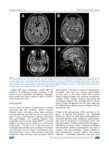
Figure 1: Magnetic resonance imaging in a patient with reversible posterior leukoencephalopathy. FLAIR axial (TR, 6160 ms; TE, 30 ms;
TI, 1100 ms; FOV 23 cm × 23 cm; matrix 256 × 192; slice thickness, 5 mm with 1 mm gap) (A); TSE T2-weighted (TR, 4500 ms; TE, 118
ms; FOV 23 cm × 23 cm; matrix 256 × 192; slice thickness, 5 mm with 1 mm gap) axial (B and C) and sagittal images (D) are shown. Figure
shows hyperintensities over both thalami, posterior limb of left internal capsule and corpus callosum on FLAIR axial image (A); hyperintense
signals are seen over bilateral parieto-occipital cortices (B) and both cerebellar hemispheres (C) on T2 axial images; hyperintensities over
corpus callosum and cerebellum are also seen in a T2 sagittal image (D) [16]
A repeat MRI done completed 2 weeks after the abnormalities in the form of areas of hypoperfusion
institution of treatment revealed resolution of the correlated well with the clinical abnormalities
lesions over the cerebellar hemispheres, brainstem, as was seen in this case. While MRI revealed
basal ganglia and cortical regions. Follow up SPECT bilateral parieto-occiptal and cerebellar hemispheric
study could not be done. changes, SPECT images revealed predominant left
hemispheric changes that correlated with the clinical
DISCUSSION picture of right hemiparesis and right gaze palsy and
the bilateral basal ganglia lesions correlated with the
This is a report of SPECT characteristics in a patient bipyramidal signs.
presenting with RPL syndrome. SPECT provides
clinically useful information about brain perfusion Central to the pathogenesis of RPL syndrome is the
and such data can especially be useful in conjunction failure of cerebral autoregulation and endothelial
with CT scan or MR imaging in diseases that cause dysfunction. When the upper limit of autoregulation is
flow abnormalities. The coupling between local exceeded arterioles dilate resulting in hyperperfusion
metabolism and blood flow allows SPECT to provide that leads to breakdown of blood-brain barrier with
indirect information about metabolism in focal areas focal transudation of fluid and petechial hemorrhages.
[1]
of the brain. [10] Hinchey et al. noted that while the Alternatively, in severe cases, it has been postulated
[1]
radiological abnormalities were symmetric, the degree that disordered cerebral autoregulation may lead to
of involvement and the clinical manifestations were reactive focal vasoconstriction, thereby resulting in
often asymmetric. Thus, in RPL syndrome where the local hypoperfusion, cytotoxic edema, and cerebral
defects are more functional than structural, SPECT infarction. Endothelial dysfunction has also been
[11]
30 Neuroimmunology and Neuroinflammation ¦ Volume 4 ¦ February 20, 2017