
Page 121 - Read Online
P. 121
Liu et al. Necroptosis and immune dysfunction in ALS
MN death by surgery in an acute manner. Because degeneration. [70,71] Furthermore, FNA can induce
of the chronic nature of ALS disease, no peak time CD4 T cell responses in both WT and SOD1 G93A mice.
+
of immune response has been defined previously. Lastly and importantly, as we mentioned in the earlier
Without a peak time, it is challenging to characterize section of “TH17 CELLS, NERVE INJURY AND
the subtle change of immune parameters. Our ALS”, FNA can “artificially” create an acute “onset”
previous studies for immune responses following of neurodegeneration and provide a predictable
nerve injury indicated that CD4 T cell responses can time frame during which to study the acute immune
+
be detected by intracellular staining for the cytokine response in mice. [64,72] Using this model, we found
production from the day 3 to day 14 post-axotomy. that FNA induced greater and prolonged immune
[64]
Such axotomy model allows us to design the best responses in draining lymph nodes of SOD1 G93A mice
measuring time point for the research and can be than WT mice, [68] suggesting Th17 cell could be a
utilized as a model tool for further study of immune potential therapeutic target for ALS treatment.
response in ALS mouse.
A HYPOTHESIZED WORKING MODEL OF ALS
While more and more genetic risks (gene mutation) for PATHOGENESIS AND FUTURE DIRECTIONS
ALS are being defined, the environmental risk factors
for ALS are relatively poorly defined. One defined Based on the literature and our data, we reason that
environmental risk is related to occupations, such as there is a cascade in the ALS disease development/
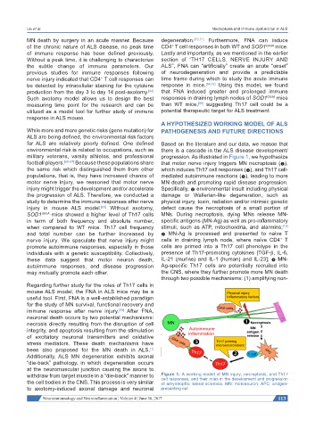
military veterans, varsity athletes, and professional progression. As illustrated in Figure 1, we hypothesize
football players. [65-67] Because these populations share that motor nerve injury triggers MN necroptosis (❶),
the same risk which distinguished them from other which induces Th17 cell responses (❷), and Th17 cell-
populations, that is, they have increased chance of mediated autoimmune reactions (❸), leading to more
motor nerve injury, we reasoned that motor nerve MN death and promoting rapid disease progression.
injury might trigger the development and/or accelerate Specifically, ❶ environmental insult including physical
the progression of ALS. Therefore, we conducted a damage or Wallerian-like degeneration, such as
study to determine the immune responses after nerve physical injury, toxin, radiation and/or intrinsic genetic
injury in mouse ALS model. [68] Without axotomy, defect cause the necroptosis of a small portion of
SOD1 G93A mice showed a higher level of Th17 cells MNs. During necroptosis, dying MNs release MN-
in term of both frequency and absolute number, specific antigens (MN-Ag) as well as pro-inflammatory
when compared to WT mice. Th17 cell frequency stimuli, such as ATP, mitochondria, and alarmins;
[18]
and total number can be further increased by ❷ MN-Ag is processed and presented to naive T
+
nerve injury. We speculate that nerve injury might cells in draining lymph node, where naïve CD4 T
promote autoimmune responses, especially in those cells are primed into a Th17 cell phenotype in the
individuals with a genetic susceptibility. Collectively, presence of Th17-promoting cytokines [TGF-β, IL-6,
these data suggest that motor neuron death, IL-21 (murine) and IL-1 (human) and IL-23]; ❸ MN-
autoimmune responses, and disease progression Ag-specific Th17 cells are potentially recruited into
may mutually promote each other. the CNS, where they further promote more MN death
through two possible mechanisms: (1) amplifying non-
Regarding further study for the roles of Th17 cells in
mouse ALS model, the FNA in ALS mice may be a
useful tool. First, FNA is a well-established paradigm
for the study of MN survival, functional recovery and
immune response after nerve injury. [69] After FNA,
neuronal death occurs by two potential mechanisms:
necrosis directly resulting from the disruption of cell
integrity, and apoptosis resulting from the stimulation
of excitatory neuronal transmitters and oxidative
stress mediators. These death mechanisms have
been also proposed for the MN death in ALS.
[1]
Additionally, ALS MN degeneration exhibits axonal
“die-back” pathology, in which degeneration occurs
at the neuromuscular junction causing the axons to
withdraw from target muscle in a “die-back” manner to Figure 1: A working model of MN injury, necroptosis, and Th17
cell responses, and their roles in the development and progression
the cell bodies in the CNS. This process is very similar of amyotrophic lateral sclerosis. MN: motoneuron; APC: antigen-
to axotomy-induced axonal damage and neuronal presenting cell
Neuroimmunology and Neuroinflammation ¦ Volume 4 ¦ June 16, 2017 113