
Page 232 - Read Online
P. 232
Menon et al. Pediatric anti-GAD antibody mediated encephalitis
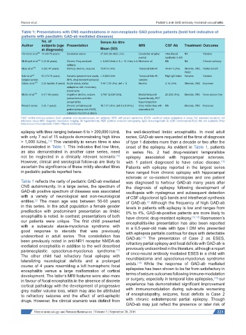
Table 1: Presentations with CNS manifestations in non-neoplastic GAD positive patients (bold font indicative of
patients with paediatric GAD-ab mediated diseases)
No. of Serum Ab titre
Author subjects (age Presentation MRI CSF Ab Treatment Outcome
at diagnosis) Mean (SD)
Honnorat et al. [6] 14 (40-70 years) Cerebellar ataxia 37,300 (30,460) U/mL Cerebellar atrophy/ Intra-thecal NA Variable
normal synthesis in 6/9
McKnight et al. [16] 5 (3-36 years) Chronic Drug resistant > 1,000 U/mL in 3; > 10 U/mL in 2 Normal in all NA NA Chronic epilepsy
epilepsy
Mata et al. [15] 2 (20, 47 years) Memory decline, seizures 72-87.5 U/mL Temporal lobe HI 46-54.1 U/mL Steroids, IVIG, Partial benefit
PLEX
[2]
Saiz et al. 50 (13-79 years) Variable (predominant ataxia, > 2,000 U/mL Temporal lobe HI, High IgG index Variable Variable
(largest series) SPS, drug resistant epilepsy) variable
[17]
Ozkan et al. 2 (9 months, 6 years) Acute ataxia, status 1.48-1.79 U/mL (ref < 1) Normal 2.16 U/mL Steroids, IVIG Improved
epilepticus with involuntary
movements
Malter et al. [16] 9 (17-66 years) Cognitive decline, seizure 1,798-12,030 U/mL Medial temporal 29-235 U/mL Steroids, IVIG None seizure free
(presentation as limbic hyperintensity, PET
encephalitis) hypometabolism
Present series 2 (3, 7 years) Chronic extratemporal 10.7-21 U/mL (ref 0-5.0 U/mL) Grey matter loss with NA Steroids, IVIG Improved
partial epilepsy with ESES, subcortical HI
subacute myoclonus ataxia
CNS: central nervous system; GAD: glutamic acid decarboxylase; Ab: antibody; SPS: stiff person syndrome; ESES: electrical status epilepticus in sleep; SD: standard deviation; ref:
reference value; MRI: magnetic resonance imaging; HI: hyperintenisty; PET: positron emission tomography; IgG: Immunoglobulin G; CSF: cerebrospinal fluid; NA: not available; IVIG:
intravenous immunoglobulin; PLEX: Plasma exchange
epilepsy with titres ranging between 6 to > 200,000 IU/mL the well-described limbic encephalitis. In most adult
with only 7 out of 15 subjects demonstrating high titres series, GAD-ab were requested at the time of diagnosis
> 1,000 IU/mL. This variability in serum titres is also of type 1 diabetes more than a decade or two after the
[12]
demonstrated in Table 1. This indicates that low titres, onset of the epilepsy. As evident in Table 1, patients
as also demonstrated in another case series, need in series No. 2 had drug-resistant temporallobe
not be neglected in a clinically relevant scenario. epilepsy associated with hippocampal sclerosis,
[13]
However, clinical and serological follow-up are likely to with 1 patient diagnosed to have celiac disease.
[15]
ascertain the significance of these mildly elevated titres Patients with epilepsy reported in the largest series
in pediatric patients reported here. have ranged from chronic epilepsy with hippocampal
sclerosis or co-existent heterotopias and one patient
Table 1 reflects the rarity of pediatric GAD-ab mediated was diagnosed to harbour GAD-ab many years after
CNS autoimmunity. In a large series, the spectrum of the diagnosis of epilepsy following development of
GAD-ab positive spectrum of diseases was associated oscillopsia with nystagmus and subsequent detection
with a variety of neurological and non-neurological of CSF oligoclonal IgG bands and intrathecal synthesis
entities. The mean age was between 50-60 years of GAD-ab. Although the frequency of high GAD-ab
[2]
[2]
in this series. In the adult population a female gender levels in patients with epilepsy is low and ranges from
predilection with predominant presentation as limbic 0% to 4%, GAD-ab-positive patients are more likely to
encephalitis is noted. In contrast, presentations of both have chronic drug-resistant epilepsy. [16,17] Rasmussen’s
our patients were unique. The first child presented encephalitis-like presentation has also been reported
with a subacute ataxia-myoclonus syndrome with in a 6.5-year-old male with type I DM who presented
good response to steroids that was previously with epilepsia partialis continua for days with detectable
undescribed in adult series. This constellation has GAD-ab. The presentation of Case 2 as ESES,
[18]
been previously noted in anti-NR1 receptor NMDA-ab refractory partial epilepsy and focal deficits with GAD-ab is
mediated encephalitis in addition to the well described previously undescribed in the literature, although a report
paraneoplastic opsoclonus-myoclonus syndrome. of onco-neural antibody mediated ESES in a child with
[14]
The other child had refractory focal epilepsy with neuroblastoma and opsoclonus-myoclonus syndrome
lateralizing neurological deficits and a prolonged exists. While the response of GAD-ab mediated
[19]
course of 4 years resembling a left hemispheric focal epilepsies has been shown to be far from satisfactory in
encephalitis versus a large malformation of cortical
development. The latter’s MRI features were also more terms of seizure outcomes following immune-modulation
[20]
in favour of focal encephalitis in the absence of discrete or surgery, especially in temporal lobe epilepsies, our
cortical pathology with the development of progressive experience has demonstrated significant improvement
grey matter volume loss, which may also be attributed with immunomodulation during sub-acute worsening
to refractory seizures and the effect of anti-epileptic of encephalopathy, seizures, focal deficits in Case 2
drugs. However, the clinical scenario was distinct from with chronic extratemporal partial epilepsy. Though
GAD-ab may just reflect the presence or later risk of
Neuroimmunology and Neuroinflammation ¦ Volume 3 ¦ September 28, 2016 223