
Page 194 - Read Online
P. 194
Feng et al. Growth cone collapse in adult sensory neurons
The results show that the EG-conditioned medium,
which contains NGF, BDNF, GDNF, and NT-3, is
neuroprotective when applied prior to or concurrently
with SEMA3A-induced collapse, and regenerative
when applied 40 min after said collapse. In previous
studies, it has been shown that when NGF, GDNF or
neuritin is applied to DRG overnight, it is protective
against SEMA3A-induced collapse. In addition,
[18]
Hansebout et al. found that EG-conditioned medium
[24]
expressed NGF, BDNF, GDNF, and NT-3 and could
induce neurite growth in cultured DRGs.
This phenomenon may be due to either neurotrophic
factor inhibition or reversal of SEMA3A-induced DRG
apoptosis. Transplanted adipose derived stem cells
[35]
into nerve conduits of rat DRG showed differentiation
of the adipose derived stem cells and the subsequent
release of NGF, BDNF, GDNF and NT-4. This was
linked with a reduction in DRG mRNA expression of
apoptotic factors Bax and caspase-3 and an increase
in expression of the anti-apoptotic factor Bcl-2. [35]
These experiments also suggest the novel finding
that NGF, BDNF, GDNF, and NT-3 are all involved
in the process of preventing or reversing SEMA3A-
induced collapse, but no individual neurotrophic factor
is essential to this process. In almost all cases, the
application of treatment medium with inhibition of
individual neurotrophic factors appeared to result in
significantly increased levels of collapse compared to
the pure EG-conditioned control medium. However,
individual inhibition did not cause full collapse, as
determined by the SEMA3A control group. This is
most apparent in the pre-treatment and co-treatment
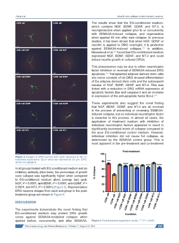
Figure 4: Images of DRG neurons from each sub-group in the co-
treatment experiment. Each white bar represents 25 μm. DRG:
dorsal root ganglion
In all groups treated with EG-conditioned medium and an
inhibitory antibody (blue bars), the percentage of growth
cone collapse was significantly higher when compared
to EG-conditioned medium alone (orange bar) (anti-
NGF, P < 0.0001; anti-BDNF, P < 0.0001; anti-GDNF, P <
0.0001; anti-NT3, P < 0.0001) [Figure 5]. Representative
DRG neurons images from each sub-group in the post-
treatment group are shown in Figure 6.
DISCUSSION
The experiments demonstrate the novel finding that
EG-conditioned medium may protect DRG growth
cones against SEMA3A-mediated collapse when
applied before, concurrently, and after SEMA3A. Figure 5: Post-treatment experiment results. ****P < 0.0001
Neuroimmunology and Neuroinflammation ¦ Volume 3 ¦ August 31, 2016 185