
Page 193 - Read Online
P. 193
Feng et al. Growth cone collapse in adult sensory neurons
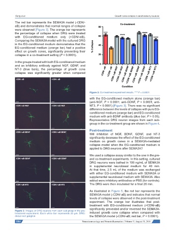
The red bar represents the SEMA3A model (-CDN/-
aB) and demonstrates that normal ranges of collapse
were observed [Figure 3]. The orange bar represents
the percentage of collapse when DRG were treated
with EG-conditioned medium only (+CDN/-aB).
Comparing the SEMA3A model with the cultured DRG
in the EG-conditioned medium demonstrates that the
EG-conditioned medium (orange bar) had a positive
effect on growth cones, significantly preventing their
collapse in a co-treatment setting (P < 0.0001).
In the groups treated with both EG-conditioned medium
and an inhibitory antibody against NGF, GDNF, and
NT-3 (blue bars), the percentage of growth cone
collapse was significantly greater when compared
Figure 3: Co-treatment experiment results. ****P < 0.0001
with the EG-conditioned medium alone (orange bar)
(anti-NGF, P < 0.0001; anti-GDNF, P < 0.0001; anti-
NT3, P < 0.0001) [Figure 3]. There was no significant
difference between the levels of collapse with pure EG-
conditioned medium (orange bar) and EG-conditioned
medium with anti-BDNF antibody (blue bar; P > 0.05).
Representative DRG neuron images from each sub-
group in the co-treatment group are shown in Figure 4.
Post-treatment
Will inhibition of NGF, BDNF, GDNF, and NT-3
significantly decrease the effect of the EG-conditioned
medium on growth cones in a SEMA3A-mediated
collapse model when the EG-conditioned medium is
applied to DRG neurons after SEMA3A?
We used a collapse assay similar to the one in the pre-
and co-treatment experiments. In this setting, cultured
DRG neurons were bathed in 100 ng/mL of SEMA3A
in supplemental neurobasal medium for 40 min.
At that time, 2.5 mL of the medium was exchanged
with either EG-conditioned medium with SEMA3A or
supplemental neurobasal medium with SEMA3A. Also
added were inhibitory antibodies or PBS (for controls).
The DRG were then incubated for a final 20 min.
As illustrated in Figure 5, the red bar represents the
SEMA3A model (-CDN/-aB) and indicates that normal
levels of collapse were observed in the post-treatment
experiment. The orange bar illustrates that post-
treatment with EG-conditioned medium (+CDN/-aB)
significantly prevented and/or reversed the SEMA3A-
Figure 2: Images of DRG neurons from each sub-group in the pre-
treatment experiment. Each white bar represents 25 μm. DRG: induced growth cone collapse when compared with
dorsal root ganglion the SEMA3A model (-CDN/-aB; red bar, P < 0.0001).
184 Neuroimmunology and Neuroinflammation ¦ Volume 3 ¦ August 31, 2016