
Page 192 - Read Online
P. 192
Feng et al. Growth cone collapse in adult sensory neurons
cone morphology was not as obvious; therefore, we
examined the stained neurons under the 40× objective
of a Leica fluorescent microscope. A minimum of 20
neurite-containing photographs were taken per slide
and the first 50 different neurons were counted per
slide in a horizontal strip manner. Growth cones were
scored as “uncollapsed” (any flattened lamellipodia
and 2 or more filopodia) or “collapsed” (bullet-
shaped neurite tip sometimes with a single filopodium
originating at the neurite tip and no lamellipodium),
as previously described. [16] Only axons that were
longer than the majority of other axons were scored;
axons that were in contact with another cell surface
were ignored. The results are represented as the
percentage of collapsed growth cones out of the total
number of growth cones counted.
Statistical analysis
Analyses were performed on combined data from
at least 3 separate experiments (n = 150) using
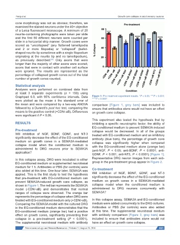
Graphpad 6.0, with 95% confidence intervals. Data Figure 1: Pre-treatment experiment results. *P < 0.05; ***P < 0.001;
were plotted as the mean ± the standard error of ****P < 0.0001
the mean and were compared by a two-way ANOVA comparison [Figure 1, grey bars] was included to
followed by a Dunnett’s post hoc test, comparing the ensure that antibodies alone would not have an effect
means to the positive control (+CDN/-aB). Differences on growth cone collapse.
were significant if P < 0.05.
This experiment also tested the hypothesis that by
RESULTS inhibiting a specific neurotrophic factor, the ability of
EG-conditioned medium to prevent SEMA3A-induced
Pre-treatment collapse would be decreased. In all of the groups
Will inhibition of NGF, BDNF, GDNF, and NT-3 treated with EG-conditioned medium and an inhibitory
significantly decrease the effect of the EG-conditioned antibody (blue bars), the percentage of growth cone
medium on growth cones in a SEMA3A-mediated collapse was significantly higher when compared
collapse model when the conditioned medium is with the EG-conditioned medium alone (orange bar)
administered to DRG neurons prior to SEMA3A (anti-NGF, P < 0.05; anti-BDNF, P < 0.0001; anti-
application?
GDNF, P < 0.001; anti-NT3, P < 0.0001) [Figure 1].
In this collapse assay, DRG were incubated in either Representative DRG neuron images from each sub-
EG-conditioned medium or supplemented neurobasal group in the pre-treatment group appear in Figure 2.
medium for 1 h. Antibodies or PBS (for controls) were
also added at this time. One hour later, SEMA3A was Co-treatment
applied. This is the first study to test the hypothesis Will inhibition of NGF, BDNF, GDNF, and NT-3
that pre-treatment with EG-conditioned medium can significantly decrease the effect of the EG-conditioned
prevent SEMA3A-induced growth cone collapse, as medium on growth cones in a SEMA3A-mediated
shown in Figure 1. The red bar represents the SEMA3A collapse model when the conditioned medium is
model (-CDN/-aB) and demonstrates that normal administered to DRG neurons concurrently with
ranges of collapse were observed. The orange bar SEMA3A?
represents the percentage of collapse when DRG were
treated with EG-conditioned medium only (+CDN/-aB). In this collapse assay, SEMA3A and EG-conditioned
Comparing the SEMA3A model with the cultured DRG medium were added concurrently to the DRG cultures.
in the EG-conditioned medium demonstrates that the Antibodies or PBS (for controls) were also added
EG-conditioned medium (orange bar) had a positive at this time. The supplemental neurobasal medium
effect on growth cones, significantly preventing their with antibody comparison [Figure 3, grey bars] was
collapse in a pre-treatment setting (P < 0.0001). included to ensure that antibodies alone would not
The supplemental neurobasal medium with antibody have an effect on growth cone collapse.
Neuroimmunology and Neuroinflammation ¦ Volume 3 ¦ August 31, 2016 183