
Page 180 - Read Online
P. 180
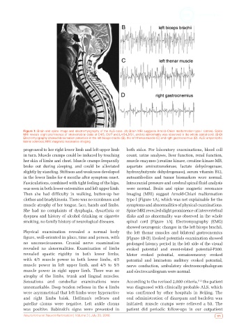
Figure 1: Brain and spine image and electromyography of the ALS case. (A) Brain MRI suggests Arnold-Chiari malformation type I (arrow). Spine
            MRI reveals slight prominence of intervertebral disks at C4/5, C6/7 and L4/5-L5/S1, and no abnormality was observed in the whole spinal cord. (B-D)
            Electromyography showed denervation potentials in the left biceps brachii (B), the left thenar muscle (C) and right gastrocnemius (D). ALS: amyotrophic
            lateral sclerosis; MRI: magnetic resonance imaging
            progressed to her right lower limb and left upper limb   both sides. For laboratory examinations, blood cell
            in turn. Muscle cramps could be induced by touching   count, urine analyses, liver function, renal function,
            her skin of limbs and chest. Muscle cramps frequently   muscle enzymes (creatine kinase, creatine kinase-MB,
            broke out during sleeping, and could be alleviated   aspartate aminotransferase, lactate dehydrogenase,
            slightly by standing. Stiffness and weakness developed   hydroxybutyrate dehydrogenase), serum vitamin B12,
            in the lower limbs for 4 months after symptom onset.   autoantibodies and tumor biomarkers were normal.
            Fasciculations, combined with tight feeling of the hips,   Intracranial pressure and cerebral spinal fluid analysis
            was seen in both lower extremities and left upper limb.   were normal. Brain and spine magnetic resonance
            Then she had difficulty in walking, button-up her   imaging (MRI) suggest Arnold-Chiari malformation
            clothes and bradykinesia. There was no numbness and   type I [Figure 1A], which was not explainable for the
            muscle atrophy of her tongue, face, hands and limbs.   symptoms and abnormalities of physical examinations.
            She had no complaints of dysphagia, dysarthria or   Spine MRI revealed slight prominence of intervertebral
            dyspnea and history of alcohol drinking or cigarette   disks and no abnormality was observed in the whole
            smoking, no family history of neurological diseases.  spinal cord [Figure 1A]. Electromyography (EMG)
                                                              showed neurogenic changes in the left biceps brachii,
            Physical examination revealed a normal body       the left thenar muscles and bilateral gastrocnemius
            figure, well-oriented in place, time and person, with   [Figure 1B-D]. Evoked potentials examination showed
            no unconsciousness. Cranial nerve examination     prolonged latency period in the left side of the visual
            revealed no abnormalities. Examination of limbs   evoked potential and event-related potential-P300.
            revealed spastic rigidity in both lower limbs,    Motor evoked potential, somatosensoroy evoked
            with 4/5 muscle power in both lower limbs, 4/5    potential  and  brainstem auditory evoked potential,
            muscle power in left upper limb, and 4/5 to 5/5   nerve conduction, ambulatory electroencephalogram
            muscle power in right upper limb. There was no    and electrocardiogram were normal.
            atrophy of the limbs, trunk and lingual muscles.
            Sensations and cerebellar examinations were       According to the revised 2,000 criteria,  the patient
                                                                                                 [12]
            unremarkable. Deep tendon reflexes in the 4 limbs   was diagnosed with clinically probable ALS, which
            were asymmetrical that left limbs were hyperactive   was confirmed by other hospitals in Beijing. The
            and right limbs brisk. Hoffman’s reflexes and     oral administration of diazepam and baclofen was
            patellar clonus were negative. Left ankle clonus   initiated; muscle cramps were relieved a bit. The
            was positive. Babinski’s signs were presented in   patient did periodic follow-ups in our outpatient
            Neuroimmunol Neuroinflammation | Volume 3 | July 20, 2016                                     171