
Page 78 - Read Online
P. 78
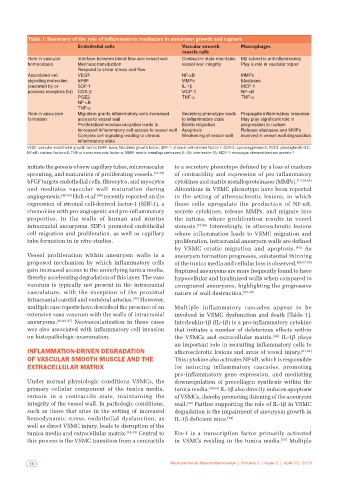
Table 1: Summary of the role of inflammatory mediators in aneurysm growth and rupture
                            Endothelial cells                    Vascular smooth      Macrophages
                                                                 muscle cells
           Role in vascular   Interface between blood flow and vessel wall  Contractile state maintains  M2 subset is antiinflammatory
           homeostasis      Mechanotransduction                  vessel wall integrity  Play a role in vascular repair
                            Respond to shear stress and flow
           Associated cell   VEGF                                NF-κB                MMPs
           signaling molecules   bFBF                            MMPs                 Elastases
           (secreted by or   SDF-1                               IL-1β                MCP-1
           possess receptors for)  COX-2                         MCP-1                NF-κB
                            PGE2                                 TNF-α                TNF-α
                            NF-κB
                            TNF-α
           Role in aneurysm   Migration grants inflammatory cells increased   Secretory phenotype leads  Propagate inflammatory response
           formation        access to vessel wall                to inflammatory state  May play significant role in
                            Proliferation/neovascularization leads to   Erratic migration  progression to rupture
                            increased inflammatory cell access to vessel wall  Apoptosis  Release elastases and MMPs
                            Complex cell signaling leading to chronic   Weakening of vessel wall  involved in vessel wall degradation
                            inflammatory state
           VEGF: vascular endothelial growth factor; bFBF: basic fibroblast growth factor; SDF‑1: stromal cell‑derived factor‑1; COX‑2: cyclooxygenase‑2; PGE2: prostaglandin E2;
           NF‑κB: nuclear factor‑κB; TNF‑α: tumor necrosis factor‑α; MMP: matrix metalloproteinases; IL‑1β: interleukin‑1β; MCP‑1: monocyte chemoattractant protein‑1

           initiate the genesis of new capillary tubes, microvascular   to a secretory phenotype defined by a loss of markers
           sprouting, and maturation of proliferating vessels. [37-39]    of contractility and expression of pro-inflammatory
           bFGF targets endothelial cells, fibrocytes, and myocytes   cytokines and matrix metalloproteinases (MMPs). [17,50-54]
           and mediates vascular wall maturation during       Alterations in VSMC phenotype have been reported
           angiogenesis. [40-43]  Hoh et al. [44]  recently reported on the   in the setting of atherosclerotic lesions, in which
           expression of stromal cell-derived factor-1 (SDF-1), a   these cells upregulate the production of NF-κB,
           chemokine with pro-angiogenic and pro-inflammatory   secrete cytokines, release MMPs, and migrate into
           properties, in the walls of human and murine       the intima, where proliferation results in vessel
           intracranial aneurysms. SDF-1 promoted endothelial   stenosis. [55,56]  Interestingly, in atherosclerotic lesions

           cell migration and proliferation, as well as capillary   where inflammation leads to VSMC migration and
           tube formation in in vitro studies.                proliferation, intracranial aneurysm walls are defined
                                                              by VSMC erratic migration and apoptosis.   [48]  As
           Vessel proliferation within aneurysm walls is a    aneurysm formation progresses, substantial thinning
           proposed mechanism by which inflammatory cells     of the tunica media and cellular loss is observed. [48,57,58]
           gain increased access to the underlying tunica media,   Ruptured aneurysms are more frequently found to have
           thereby accelerating degradation of this layer. The vaso   hypocellular and hyalinized walls when compared to
           vasorum is typically not present in the intracranial   unruptured aneurysms, highlighting the progressive
           vasculature,  with  the  exception  of  the  proximal   nature of wall destruction. [59,60]
           intracranial carotid and vertebral arteries. [45]  However,
           multiple case reports have described the presence of an   Multiple inflammatory cascades appear to be
           extensive vaso vasorum with the walls of intracranial   involved in VSMC dysfunction and death [Table 1].
           aneurysms. [44,46,47]  Neovascularization in these cases   Interleukin-1β (IL-1β) is a pro-inflammatory cytokine
           was also associated with inflammatory cell invasion   that initiates a number of deleterious effects within
           on histopathologic examination.                    the VSMCs and extracellular matrix. [48]  IL-1β plays
                                                              an important role in recruiting inflammatory cells to
           INFLAMMATION‑DRIVEN DEGRADATION                    atherosclerotic lesions and areas of vessel injury. [61,62]
           OF VASCULAR SMOOTH MUSCLE AND THE                  This cytokine also activates NF-κB, which is responsible
           EXTRACELLULAR MATRIX                               for inducing inflammatory cascades, promoting
                                                              pro-inflammatory gene expression, and mediating
           Under normal physiologic conditions VSMCs, the     downregulation of procollagen synthesis within the
           primary cellular component of the tunica media,    tunica media. [48,63]  IL-1β also directly induces apoptosis
           remain  in  a  contractile  state,  maintaining  the   of VSMCs, thereby promoting thinning of the aneurysm
           integrity of the vessel wall. In pathologic conditions,   wall. [48]  Further supporting the role of IL-1β in VSMC
           such as those that arise in the setting of increased   degradation is the impairment of aneurysm growth in
           hemodynamic stress, endothelial dysfunction, as    IL-1β deficient mice. [64]
           well as direct VSMC injury, leads to disruption of the
           tunica media and extracellular matrix. [48,49]  Central to   Ets-1 is  a  transcription factor  primarily  activated
           this process is the VSMC transition from a contractile   in VSMCs residing in the tunica media. [65]  Multiple


            70                                               Neuroimmunol Neuroinflammation | Volume 2 | Issue 2 | April 15, 2015