
Page 300 - Read Online
P. 300
laboratory tests about thyroid function, autoimmune occlusive site and predilation was performed with
antibodies, and vasculitis indicators were all normal. a balloon (2.5 mm × 15 mm) at 6 atm. Then, the
stent (4 mm × 40 mm Xpert Stent System) was deployed
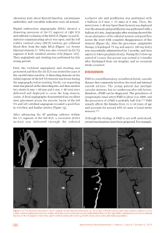
Digital subtraction angiography (DSA) showed a over the stenosis and postdilation was performed with a
dissecting aneurysm of the C1 segment of right ICA balloon at 6 atm. Angiography after stenting showed the
and subtotal occlusion of the left ICA [Figure 1a and b]. revascularization of the subtotal stenosis and good flow
Anterior communicating artery was open, and the left across the stent with complete disappearance of this
middle cerebral artery (MCA) territory got collateral stenosis [Figure 1h]. After the procedure, antiplatelet
blood flow from the right MCA [Figure 1c]. Severe therapy (clopidogrel 75 mg and aspirin 100 mg daily)
bilateral stenosis (> 70%) was also revealed on the V2 was sequentially administered for 3 months, and then
segment of both vertebral arteries (VA) [Figure 1d‑f]. aspirin is taken prophylactically. During the follow‑up
Then angioplasty and stenting was performed for this period of 3 years, this patient was normal at 3 months
young patient. after discharged from our hospital, and no recurrent
stroke occurred.
First, the vertebral angioplasty and stenting was
performed and then the left ICA was stented because of DISCUSSION
the carotid sinus reaction. A dissecting stenosis on the
initial segment of the left VA stenosis was found during FMD is a noninflammatory, nonatherosclerotic vascular
the angiography before stenting. Firstly, one expanding disease that commonly involves the renal and internal
stent was placed on the dissecting site, and then another carotid arteries. The young patient has multiple
two stents (4 mm × 60 mm and 4 mm × 40 mm) were vascular stenoses, but no cerebrovascular risk factors;
delivered and deployed to cover the long stenotic therefore, cFMD can be diagnosed. The prevalence of
lesion. A final angiography demonstrated an excellent symptomatic renal artery FMD is about 4 in 1000, and
stent placement across the stenotic lesion of the left the prevalence of cFMD is probably half that. [1‑4] FMD
VA and left vertebral angiogram revealed a good flow usually affects the females from 15 to 50 years of age
in vertebral and basilar arteries [Figure 1g]. and accounts for around 10% of cases of renal artery
stenosis. [1‑4]
After advancing the 8F guiding catheter within
the C1 segment of the left ICA, a microwire (0.014 Although the etiology of FMD is not well understood,
inches) was delivered through the subtotal several mechanisms have been proposed. For example,
a b c d
e f g h
Figure 1: (a) The dissecting aneurysm on the C1 segment of right internal carotid artery; (b) “rat tail sign” of the left internal carotid artery before predilation; (c) the
anterior communicating artery was open and the left middle cerebral artery territory got collateral blood flow from the right middle cerebral artery; (d‑f) severe bilateral
stenosis on the V2 segment of both VAs; (g) left vertebral arteries after stenting; (h) left internal carotid artery after balloon predilation
292 Neuroimmunol Neuroinflammation | Volume 2 | Issue 4 | October 15, 2015 Neuroimmunol Neuroinflammation | Volume 2 | Issue 4 | October 15, 2015 293