
Page 297 - Read Online
P. 297
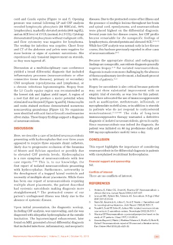
cord and Cauda equina [Figure 1e and f]. Opening   diseases. Due to the protracted course of her illness and
           pressure was normal following LP and CSF analysis   the presence of multiple lesions throughout her brain
           revealed lymphocytic pleocytosis (88 WBCs/uL, 94%   and spinal cord, ependymoma, and neurosarcoidosis
           lymphocytes), markedly elevated protein (868 mg/dL),   were placed highest on the differential diagnosis.
           and an ACE level of 15 U/L (normal, 0‑2.5 U/L). Cytology   Several years into her disease course, her CSF profile
           demonstrated lymphocytosis without malignant cells,   became remarkable for the nonspecific findings of
           and  flow  cytometry  was  negative  for  lymphoma.   lymphocytosis, elevated protein and elevated ACE. [5,12,13]
           The workup for infection was negative. Chest X‑ray   While her CSF analysis was normal early in her disease
           and CT of the abdomen and pelvis were negative for   course, this has been previously reported in other cases
           mass  lesions  or  signs  of systemic sarcoidosis. She   of neurosarcoidosis. [6,7]
           experienced only transient improvement on steroids,
           so they were tapered off.                          Because the appropriate clinical and radiographic
                                                              findings are nonspecific, sarcoidosis diagnosis generally
           Discussion at a multidisciplinary case conference   requires biopsy. [1‑3]  For isolated neurosarcoidosis,
           yielded a broad differential diagnosis that included   diagnosis is made even more challenging by the absence
           inflammatory processes  (neurosarcoidosis or other   of thoracopulmonary involvement, a hallmark present
           connective tissue  diseases), primary  or secondary   in 90% of patients. [1,2]
           CNS neoplasm  (ependymoma and lymphoma), or
           a chronic infectious leptomeningitis. Biopsy from   Biopsy for sarcoidosis is also critical because patients
           the L3  Cauda equina region was recommended as     may not show substantial improvement with an
           the lowest risk and highest yield target. A region of   empiric trial of steroids, as was true for this patient.
           a root that did not produce a motor potential when   Many have advocated the use of immunosuppressants
           stimulated was biopsied [Figure 1g and h]. Hematoxylin   such as azathioprine, methotrexate, infliximab, or
           and eosin stained sections demonstrated numerous   mycophenolate mofetil alone, or in addition to steroids
           noncaseating granulomas [Figure 1i]. No organisms   in patients who do not respond to corticosteroid
           were identified with acid‑fast or Grocott’s methenamine   monotreatment. [1,2,14‑16]   The  risks  of  long‑term
           silver stains. These biopsy findings support a diagnosis   immunosuppressive therapy warranted a definitive
           of neurosarcoidosis.                               diagnosis of isolated neurosarcoidosis, given its rarity.
                                                              Since neurosarcoidosis was indeed the diagnosis, the
           DISCUSSION                                         patient was initiated on 80 mg prednisone daily and
                                                              500 mg mycophenolate mofetil twice a day.
           Here, we describe a case of isolated neurosarcoidosis
           presenting with hydrocephalus that over three years   CONCLUSION
           appeared to require three separate shunt catheters,
           likely due to progressive occlusion of the foramina   This report highlights the importance of considering
           of  Monro  and  Sylvian  aqueduct  or  possibly  due   neurosarcoidosis in the differential diagnosis in patients
           to elevated CSF protein levels. Hydrocephalus      with unexplained recalcitrant hydrocephalus.
           is a rare symptom of neurosarcoidosis with few
           case reports. [7‑10]  This is, to our knowledge, the   Financial support and sponsorship
           first report of isolated neurosarcoidosis presenting   Nil.
           with hydrocephalus.  Furthermore,  noteworthy  is
           the development of a trapped lateral ventricle and   Conflicts of interest
           necessity of multiple shunt placements. While there   There are no conflicts of interest.
           has been one report of neurosarcoidosis requiring
           multiple shunt placements, the patient described   REFERENCES
           had systemic sarcoidosis making diagnosis more     1.   Hoitsma E, Faber CG, Drent M, Sharma OP. Neurosarcoidosis: a
                           [8]
           straightforward.  The protracted course of our         clinical dilemma. Lancet Neurol 2004;3:397‑407.
           patient’s undiagnosed illness was likely due to the   2.   Iannuzzi MC, Rybicki BA, Teirstein AS. Sarcoidosis. N Engl J Med
           absence of systemic disease.                           2007;357:2153‑65.
                                                              3.   Stern BJ, Krumholz A, Johns C, Scott P, Nissim J. Sarcoidosis and
                                                                  its neurological manifestation. Arch Neurol 1985;42:909‑17.
           Upon initial presentation, the diagnostic workup,   4.   Nozaki K, Scott TF, Sohn M, Judson MA. Isolated neurosarcoidosis:
           including CSF analysis, was unrevealing, and she was   case series in 2 sarcoidosis centers. Neurologist 2012;18:373‑7.
           diagnosed with idiopathic hydrocephalus at the outside   5.   Sharma OP. Neurosarcoidosis: a personal perspective based on the
           institution. The leptomeningeal enhancement, later     study of 37 patients. Chest 1997;112:220‑8.
           found on MRI, generated a broad differential diagnosis   6.   Gascón‑Bayarri J, Mañá J, Martínez‑Yélamos S, Murillo O, Reñé R,
                                                                  Rubio F. Neurosarcoidosis: report of 30 cases and a literature survey.
           that included infectious, inflammatory, and neoplastic   Eur J Intern Med 2011;22:e125‑32.


   288  Neuroimmunol Neuroinflammation | Volume 2 | Issue 4 | October 15, 2015  Neuroimmunol Neuroinflammation | Volume 2 | Issue 4 | October 15, 2015   289