
Page 771 - Read Online
P. 771
de Divitiis et al. Mini-invasive Surg 2020;4:75 I http://dx.doi.org/10.20517/2574-1225.2020.66 Page 7 of 10
A B
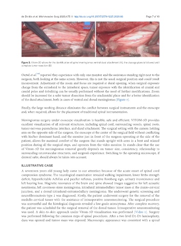
Figure 3. Vitom-3D allows for the identification of spinal meningiomas ventral dural attachment (A); the cleavage plane is followed until
complete tumor resection (B)
[39]
Oertel et al. reported they experience with only one monitor and the assistance standing right next to the
surgeon, both looking at the same screen. However, this is not the usual surgical position and could result
inconvenient. Adjustment of the zoom and focus are required at dural opening, when surgical exposure
change from the extradural to the intradural space; tumor exposure with the identification of cranial and
caudal poles and debulking can be usually performed without the need of further modifications. Zoom
should be increased for a safer tumor dissection from the arachnoidal plane and for a better identification
of the dural attachment, both in cases of ventral and dorsal meningiomas [Figure 3].
Finally, the large working distance eliminates the conflict between surgical instruments and the exoscope
and, when required, allows for the placement of traditional spinal instrumentation.
Meningiomas surgery under exoscope visualization is feasible, safe and efficient. VITOM-3D provides
excellent visualization of all relevant structures, including spinal cord, surrounding vessels, spinal roots,
tumor-nervous parenchyma interface, and dural attachment. The surgical setting with the camera holding
arm on the opposite side of the surgeon, the exoscope at the center of the surgical field without conflicting
with his/her dominant hand, and the monitor just in front of the surgeon operating at the side of the
patient, allows for maximal comfort of the surgeon that stands upright with arms in a bent and relaxed
position during all the surgical steps, and operates from the video monitor. In stands clear that the use
of Vitom-3D for meningiomas removal greatly depends on tumor size, consistency, relationship to
surrounding neurovascular structures, and surgeon’s experience. Switching to the operating microscope, if
deemed safer, should always be taken into account.
ILLUSTRATIVE CASE
A seventeen years old young lady came to our attention because of the acute onset of spinal cord
compression syndrome. The neurological examination revealed walking impairment, lower limbs strength
deficit, hyperelicitable Achilles and patellar reflexes, positive Romberg sign, urinary incontinence and
left hearing loss. Magnetic resonance of the brain and spine showed images suggestive for left acoustic
neurinoma, left cavernous sinus meningioma, intradural intramedullary tumor mass at the cranio-cervical
junction, and a dorsal intradural-extramedullary meningioma. She underwent genetic screening and
neurofibromatosis type 2 was diagnosed. Firstly, the patient underwent surgery for the removal of the
medullo-cervical tumor with the assistance of intraoperative neuromonitoring. The surgical procedure
was uneventful and the histological diagnosis revealed a low-grade astrocytoma. After complete recovery,
the patient was scheduled for the surgical removal of the dorsal tumor. Intraoperative neuromonitoring
was used. A skin-to-skin approach under Vitom-3D visualization was performed [Video 1]. Surgery
was performed following the common steps of spinal procedures. After a two level D2-D3 laminoplasty,
dura was opened and tumor mass was exposed. Macroscopic appearance was consistent with a spinal