
Page 768 - Read Online
P. 768
Page 4 of 10 de Divitiis et al. Mini-invasive Surg 2020;4:75 I http://dx.doi.org/10.20517/2574-1225.2020.66
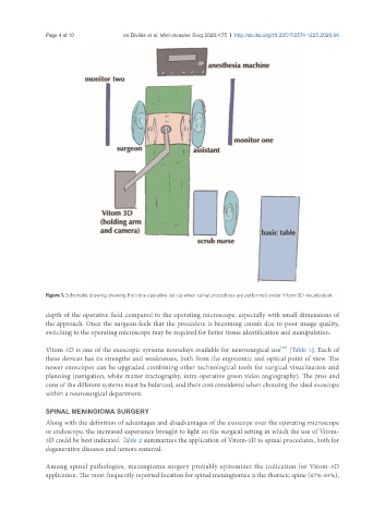
Figure 1. Schematic drawing showing the intra-operative set-up when spinal procedures are performed under Vitom-3D visualization
depth of the operative field compared to the operating microscope, especially with small dimensions of
the approach. Once the surgeon feels that the procedure is becoming unsafe due to poor image quality,
switching to the operating microscope may be required for better tissue identification and manipulation.
[55]
Vitom-3D is one of the exoscopic systems nowadays available for neurosurgical use [Table 1]. Each of
these devices has its strengths and weaknesses, both from the ergonomic and optical point of view. The
newer exoscopes can be upgraded combining other technological tools for surgical visualization and
planning (navigation, white matter tractography, intra-operative green video angiography). The pros and
cons of the different systems must be balanced, and their cost considered when choosing the ideal exoscope
within a neurosurgical department.
SPINAL MENINGIOMA SURGERY
Along with the definition of advantages and disadvantages of the exoscope over the operating microscope
or endoscope, the increased experience brought to light on the surgical setting in which the use of Vitom-
3D could be best indicated. Table 2 summarizes the application of Vitom-3D to spinal procedures, both for
degenerative diseases and tumors removal.
Among spinal pathologies, meningioma surgery probably epitomizes the indication for Vitom-3D
application. The most frequently reported location for spinal meningiomas is the thoracic spine (67%-84%),