
Page 770 - Read Online
P. 770
Page 6 of 10 de Divitiis et al. Mini-invasive Surg 2020;4:75 I http://dx.doi.org/10.20517/2574-1225.2020.66
A B
C D
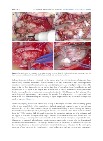
Figure 2. Two levels dorsal laminotomy is performed under visualization of Vitom-3D (A, B); after focus and zoom adjustment, the
tumor is exposed and dissected from arachnoidal adherences (C, D) until total removal is achieved
followed by the cervical spine (14%-27%), and the lumbar spine (2%-14%). At the time of diagnosis, these
tumors rarely extend for more than 3 laminae, because of the early occurrence of signs and symptoms of
spinal cord compression in their natural history. Considering the above, the optical properties of Vitom-3D,
in particular the focal length of 25-60 cm and the large field of view, allow for excellent illumination and
magnification of the depth of the surgical field, more in cases of cervical and thoracic meningiomas than
lumbar tumors, where the operative field is deeper. Positioning the VITOM camera at the beginning of the
surgical approach approximately 35-40 cm above the operative field, all procedures can be performed with
the minimal need for repositioning and refocusing at higher magnifications, due to the quite homogeneous
depth of exposure [Figure 2].
In this way, ongoing video documentation step-by-step of the surgical procedure with outstanding quality
of the images is available for all the surgical room staff and educational purpose. In cases of meningiomas
extending for more than three laminae, exoscope adjustments would likely be more often required. During
the extradural steps of the procedure, the surgeon, if wished, could operate under direct vision rather than
from the VITOM monitor. Still, it is worth to consider the exoscope a teaching tool of great impact and
we suggest its utilization during the whole surgery. Besides, the use of the VITOM from skin incision may
help in reducing the learning curve that is associated to the introduction on any new surgical instrument.
Fluoroscopy is commonly adopted during meningiomas surgery in order to safely tailor the dimension of
the approach to tumor extension. The exoscope doesn’t need to be transitioned in and out of the operative
field during placement of fluoroscopy, which may contribute to increased efficiency. On the counterpart,
the need of two monitors for spinal surgery could reduce the working environment ergonomic.