
Page 767 - Read Online
P. 767
de Divitiis et al. Mini-invasive Surg 2020;4:75 I http://dx.doi.org/10.20517/2574-1225.2020.66 Page 3 of 10
[22]
In 2008, Mamelak et al. reported the initial use of this novel tool in an animal model. The initial
impression was that the exoscope had the potential for widespread application for human microsurgical
procedures owing adequate image resolution, magnification, and easy and intuitive manipulation. Soon
after the initial report, its clinical use has been tested in many surgical disciplines including vascular
and cardiac surgery, ENT, hepatic surgery, and neurosurgery [23,26,29-34] . The outstanding quality of images
was largely confirmed and the exoscope earned the right to be seen as another visualization tool in the
armamentarium of the contemporary neurosurgeon. The first clinical series of patients undergoing surgery
with the aim of the exoscope consisted of technically less demanding neurosurgical procedures for which
it was felt that trial use of this device could not potentially affect surgical outcomes. That lack of 3D
visualization was a concern to many surgeons and this drawback was mentioned frequently in preliminary
studies [24,35-38] . It took about nine years to further evolve the 2D visualization and introduce the first 3D
exoscopic visualization system [39,40] . In the following years, the 3D exoscope has been used to perform
various technical more demanding neurosurgical procedures, even in pediatric cases, including treatment
of cerebrovascular disorders (i.e., aneurysm clipping and bypass surgery), degenerative spinal disorders,
and resection of cranial and spinal tumors [41-50] .
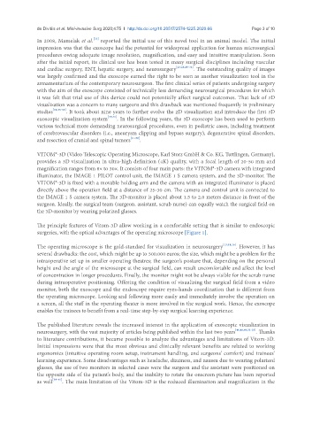
VITOM®-3D (Video Telescopic Operating Microscope, Karl Storz GmbH & Co. KG, Tuttlingen, Germany),
provides a 3D visualization in ultra-high definition (4K) quality, with a focal length of 20-50 mm and
magnification ranges from 8× to 30×. It consists of four main parts: the VITOM®-3D camera with integrated
illuminator, the IMAGE 1 PILOT control unit, the IMAGE 1 S camera system, and the 3D-monitor. The
VITOM®-3D is fixed with a movable holding arm and the camera with an integrated illuminator is placed
directly above the operation field at a distance of 25-30 cm. The camera and control unit is connected to
the IMAGE 1 S camera system. The 3D-monitor is placed about 1.5 to 2.0 meters distance in front of the
surgeon. Ideally, the surgical team (surgeon, assistant, scrub nurse) can equally watch the surgical field on
the 3D-monitor by wearing polarized glasses.
The principle features of Vitom-3D allow working in a comfortable setting that is similar to endoscopic
surgeries, with the optical advantages of the operating microscope [Figure 1].
The operating microscope is the gold-standard for visualization in neurosurgery [13,14,16] . However, it has
several drawbacks: the cost, which might be up to 500.000 euros; the size, which might be a problem for the
intraoperative set up in smaller operating theatres; the surgeon’s posture that, depending on the personal
height and the angle of the microscope at the surgical field, can result uncomfortable and affect the level
of concentration in longer procedures. Finally, the monitor might not be always visible for the scrub nurse
during intraoperative positioning. Offering the condition of visualizing the surgical field from a video
monitor, both the exoscope and the endoscope require eyes-hands coordination that is different from
the operating microscope. Looking and following more easily and immediately involve the operation on
a screen, all the staff in the operating theater is more involved in the surgical work. Hence, the exoscope
enables the trainees to benefit from a real-time step-by-step surgical learning experience.
The published literature reveals the increased interest in the application of exoscopic visualization in
neurosurgery, with the vast majority of articles being published within the last two years [46,48,49,51-55] . Thanks
to literature contributions, it became possible to analyze the advantages and limitations of Vitom-3D.
Initial impressions were that the most obvious and clinically relevant benefits are related to working
ergonomics (intuitive operating room setup, instrument handling, and surgeons’ comfort) and trainees’
learning experience. Some disadvantages such as headache, dizziness, and nausea due to wearing polarized
glasses, the use of two monitors in selected cases were the surgeon and the assistant were positioned on
the opposite side of the patient’s body, and the inability to rotate the onscreen picture has been reported
as well [39-41] . The main limitation of the Vitom-3D is the reduced illumination and magnification in the