
Page 498 - Read Online
P. 498
Cannata et al. Mini-invasive Surg 2020;4:53 I http://dx.doi.org/10.20517/2574-1225.2020.41 Page 5 of 17
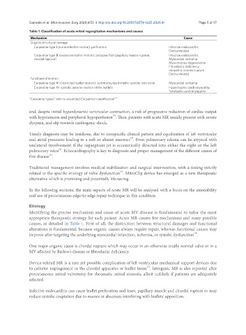
Table 1. Classification of acute mitral regurgitation mechanisms and causes
Mechanism Cause
Organic/structural damage
Carpentier type I (normal leaflet motion): perforation Infective endocarditis
Device-related
Carpentier type II (excessive leaflet motion): prolapse/flail (papillary muscle rupture, Infective endocarditis
chordal rupture) Myocardial ischemia
Myxomatous degeneration
Fibroelastic deficiency
Idiopathic chordal rupture
Device-related
Functional alteration
Carpentier type III (restricted leaflet motion): symmetric/asymmetric systolic restriction Myocardial ischemia
Carpentier type IV: systolic anterior motion of the leaflets Hypertrophic cardiomyopathy
Takotsubo cardiomyopathy
[3]
“Carpentier types” refer to expanded Carpentier classification
and, despite initial hyperdynamic ventricular contraction, a risk of progressive reduction of cardiac output
[4]
with hypotension and peripheral hypoperfusion . Thus, patients with acute MR usually present with severe
dyspnea, and slip towards cardiogenic shock.
Timely diagnosis may be insidious, due to nonspecific clinical pattern and equalization of left ventricular
[2]
and atrial pressures leading to a soft or absent murmur . Even pulmonary edema can be atypical with
unilateral involvement if the regurgitant jet is eccentrically directed into either the right or the left
[2]
pulmonary veins . Echocardiography is key to diagnosis and proper management of the different causes of
this disease .
[5]
Traditional management involves medical stabilization and surgical intervention, with a timing strictly
[6]
related to the specific etiology of valve dysfunction . MitraClip device has emerged as a new therapeutic
alternative which is promising and potentially life-saving.
In the following sections, the main aspects of acute MR will be analysed with a focus on the amenability
and use of percutaneous edge-to-edge repair technique in this condition.
Etiology
Identifying the precise mechanism and cause of acute MV disease is fundamental to tailor the most
appropriate therapeutic strategy for each patient. Acute MR counts few mechanisms and many possible
causes, as detailed in Table 1. First of all, the distinction between structural damages and functional
alterations is fundamental, because organic causes always require repair, whereas functional causes may
improve after targeting the underlying myocardial infarction, ischemia, or systolic dysfunction .
[5]
One major organic cause is chordal rupture which may occur in an otherwise totally normal valve or in a
MV affected by Barlow’s disease or fibroelastic deficiency.
Device-related MR is a rare yet possible complication of left ventricular mechanical support devices due
to catheter impingement in the chordal apparatus or leaflet tissue . Iatrogenic MR is also reported after
[7]
percutaneous mitral valvotomy for rheumatic mitral stenosis, albeit unlikely if patients are adequately
selected.
Infective endocarditis can cause leaflet perforation and tears, papillary muscle and chordal rupture or may
reduce systolic coaptation due to masses or abscesses interfering with leaflets’ apposition.