
Page 496 - Read Online
P. 496
Cannata et al. Mini-invasive Surg 2020;4:53 I http://dx.doi.org/10.20517/2574-1225.2020.41 Page 3 of 17
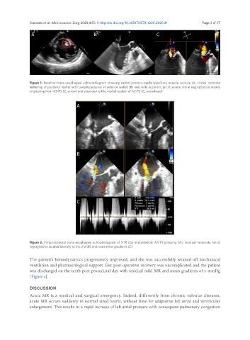
A B C
Figure 1. Baseline trans-esophageal echocardiogram showing partial postero-medial papillary muscle rupture (A, circle), extreme
tethering of posterior leaflet with pseudoprolapse of anterior leaflet (B) and wide eccentric jet of severe mitral regurgitation mainly
originating from A3-P3 (C, arrow) and extended to the medial section of A2-P2 (C, arrowhead)
A
B
C
Figure 2. Intraprocedural trans-esophageal echocardiogram of XTR clip implantation: A3-P3 grasping (A), residual moderate mitral
regurgitation located laterally to the clip (B) and transmitral gradients (C)
The patient’s hemodynamics progressively improved, and she was successfully weaned off mechanical
ventilation and pharmacological support. Her post-operative recovery was uncomplicated and the patient
was discharged on the tenth post-procedural day with residual mild MR and mean gradients of 5 mmHg
[Figure 4].
DISCUSSION
Acute MR is a medical and surgical emergency. Indeed, differently from chronic valvular diseases,
acute MR occurs suddenly in normal sized hearts, without time for adaptative left atrial and ventricular
enlargement. This results in a rapid increase of left atrial pressure with consequent pulmonary congestion