
Page 312 - Read Online
P. 312
Lutter et al. Mini-invasive Surg 2020;4:36 I http://dx.doi.org/10.20517/2574-1225.2020.15 Page 5 of 6
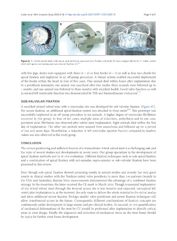
Figure 5. A: mitral valved stent with apical and additional supra-annular fixation elements; B: hook shaped elements; C: mitral valved
stent with apical and subsequent sub-valvular fixation rim [13]
with five pigs, stents were equipped with three (n = 2) or four hooks (n = 3) as well as four neo-chords for
apical fixation and implanted in an off-pump procedure. A thread system enabled successful deployment
of the hooks within the heart in four of five cases. One animal died within hours after implantation due
to a prosthesis mismatch; one animal was sacrificed after two weeks; three animals were followed up to
1 month; and one animal was followed to three months with excellent health. Good valve function as well
[11]
as normal left ventricular function was demonstrated by TEE and haemodynamic evaluation .
SUB-VALVULAR FIXATION
A modified nitinol valved stent with a ventricular rim was developed for sub-valvular fixation [Figure 5C].
[11]
For secure fixation, an additional apical fixation system was attached to these stents . This prototype was
successfully implanted in an off-pump procedure in ten animals. A higher degree of ventricular fibrillation
occurred in this group. In four of ten cases, multiple areas of infarction, arrhythmia and in one case,
persistent atrial fibrillation was observed after valved stent implantation. Eight animals died within the first
day of implantation. The other two animals were weaned from anaesthesia and followed up for a period
of two and seven days. Nevertheless, a reduction in left ventricular ejection fraction compared to baseline
values was also observed in this study group.
CONCLUSION
The correct positioning and sufficient fixation of a transcatheter mitral valved stent is a challenging task and
the topic of several studies and developments in recent years. Our group specializes in the development of
apical fixation methods and its in vivo evaluation. Different fixation techniques such as sole apical fixation
and a combination of apical fixation with sub-annular, supra-annular or sub-valvular fixation have been
presented in this review.
Even though sole apical fixation showed promising results in animal studies and already has very good
results in clinical studies with the Tendyne mitral valve prosthesis in more than 150 patients (mainly in
the USA and Australia), fixation force measurements demonstrated the advantage of a combined fixation
strategy. In the meantime, the latter received the CE mark in March 2020. Though transseptal implantation
of the mitral valved stent through the femoral access site is less invasive and expected, transapical left
ventricular implantation is, at the moment, the only route to deliver the whole material to the mitral anulus
and allow additional secure fixation. Perhaps smaller valve prostheses and newer fixation techniques will
allow transfemoral access in the future. Consequently, different combinations of fixation concepts are
continuously under development in large animal and pre-clinical studies. To succeed, in vivo quantification
of mechanical deformations of the stent by CT should be performed after implantation to identify critical
areas in stent design. Finally, the alignment and reduction of mechanical stress on the stent frame should
be topics for further stent frame development.