
Page 309 - Read Online
P. 309
Page 2 of 6 Lutter et al. Mini-invasive Surg 2020;4:36 I http://dx.doi.org/10.20517/2574-1225.2020.15
operable. Due to the advanced age and severe comorbidities, approximately 49% of patients suffering from
[1]
severe and symptomatic mitral valve insufficiency are not candidates for open-heart surgery .
Challenges in the development process of such mitral valved stents include: high pressure within the
left heart chamber, secure fixation to the complex anatomy of the mitral valve apparatus, absence of left
ventricular outflow tract obstruction, and a tight seal to prevent the occurrence of paravalvular leakage after
valved stent implantation. Due to very high pressures within the left ventricle, which act on the closed mitral
valve, the development of strong systolic fixation of the device to prevent migration into the left atrium is of
particular importance during the research and developmental process.
TETHERED APICAL FIXATION (LUTTER VALVE)
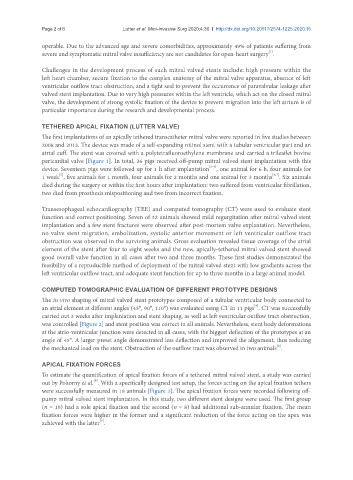
The first implantations of an apically tethered transcatheter mitral valve were reported in five studies between
2008 and 2013. The device was made of a self-expanding nitinol stent with a tubular ventricular part and an
atrial cuff. The stent was covered with a polytetrafluoroethylene membrane and carried a trileaflet bovine
pericardial valve [Figure 1]. In total, 36 pigs received off-pump mitral valved stent implantation with this
[2-4]
device. Seventeen pigs were followed up for 1 h after implantation , one animal for 6 h, four animals for
[6,7]
[5]
1 week , five animals for 1 month, four animals for 2 months and one animal for 3 months . Six animals
died during the surgery or within the first hours after implantation: two suffered from ventricular fibrillation,
two died from prosthesis mispositioning and two from incorrect fixation.
Transesophageal echocardiography (TEE) and computed tomography (CT) were used to evaluate stent
function and correct positioning. Seven of 32 animals showed mild regurgitation after mitral valved stent
implantation and a few stent fractures were observed after post-mortem valve explantation. Nevertheless,
no valve stent migration, embolization, systolic anterior movement or left ventricular outflow tract
obstruction was observed in the surviving animals. Gross evaluation revealed tissue coverage of the atrial
element of the stent after four to eight weeks and the new, apically-tethered mitral valved stent showed
good overall valve function in all cases after two and three months. These first studies demonstrated the
feasibility of a reproducible method of deployment of the mitral valved stent with low gradients across the
left ventricular outflow tract, and adequate stent function for up to three months in a large animal model.
COMPUTED TOMOGRAPHIC EVALUATION OF DIFFERENT PROTOTYPE DESIGNS
The in vivo shaping of mitral valved stent prototypes composed of a tubular ventricular body connected to
[8]
an atrial element at different angles (45°, 90°, 110°) was evaluated using CT in 11 pigs . CT was successfully
carried out 3 weeks after implantation and stent shaping, as well as left ventricular outflow tract obstruction,
was controlled [Figure 2] and stent position was correct in all animals. Nevertheless, stent body deformations
at the atrio-ventricular junction were detected in all cases, with the biggest deflection of the prototypes at an
angle of 45°. A larger preset angle demonstrated less deflection and improved the alignment, thus reducing
[8]
the mechanical load on the stent. Obstruction of the outflow tract was observed in two animals .
APICAL FIXATION FORCES
To estimate the quantification of apical fixation forces of a tethered mitral valved stent, a study was carried
[9]
out by Pokorny et al. . With a specifically designed test setup, the forces acting on the apical fixation tethers
were successfully measured in 18 animals [Figure 3]. The apical fixation forces were recorded following off-
pump mitral valved stent implantation. In this study, two different stent designs were used. The first group
(n = 10) had a sole apical fixation and the second (n = 8) had additional sub-annular fixation. The mean
fixation forces were higher in the former and a significant reduction of the force acting on the apex was
[9]
achieved with the latter .