
Page 310 - Read Online
P. 310
Lutter et al. Mini-invasive Surg 2020;4:36 I http://dx.doi.org/10.20517/2574-1225.2020.15 Page 3 of 6
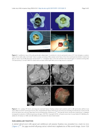
Figure 1. A: ventricular view of the atrioventricular valved stent. It consists of a bovine pericardial valve of 27 mm diameter, a custom-
made nitinol stent, and a ventricular fixation system consisting of the annular radial force of the nitinol stent and four tethers fixed at the
[3]
apex; B: atrial view of the prototype valved stent ; C: ventricular view of the new refined mitral valved stent; D: operative setting after
ministernotomy (2 inches) with this valved stent and apex of the heart with purse-string sutures is exposed [6]
Figure 2. A-C: cardiac CT short- and long-axis standard views showing correct stent position and no left ventricular outflow tract
obstruction of the 110° prototype 1 month after implantation; D: three-dimensional reconstruction showing the nitinol stent frame and left
[8]
atrial and ventricular volumes. Reprinted from EuroIntervention, Pokorny et al. , Transapical mitral valved stent implantation: computed
tomographic evaluation of different prototype designs, 948-955. Copyright (2015), with permission from Europa Digital & Publishing. Ao:
aorta; LA: left atrium; LV: left ventricle (white arrow indicates the nitinol stent frame)
SUB-ANNULAR FIXATION
A mitral valved stent with apical and additional sub-annular fixation was presented in a study in 2016
[10]
[Figure 4] . Ten pigs received off-pump mitral valved stent implantation of this novel design. Acute TEE