Page 317 - Read Online
P. 317
Page 4 of 7                                     Mammana et al. Mini-invasive Surg 2020;4:37  I  http://dx.doi.org/10.20517/2574-1225.2020.24


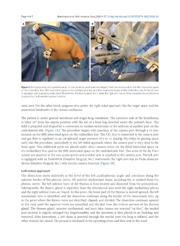
               Figure 1. Port positioning and operative setup. A: the patient is positioned and draped. Ports are introduced on the fifth intercostal space
               on the midaxillary line, fifth intercostal space on the midclavicular line, and third intercostal space on the midaxillary line; B: the left arm
               is equipped with a grasping instrument (EndoWrist, Intuitive Surgical, Inc.), while the right arm has an Endo-dissector device (Intuitive
               Surgical, Inc.) with electric cautery function

               vena cava. On the other hand, surgeons who prefer the right-sided approach like the larger space and the
               anatomical landmarks of the venous confluence.

               The patient is under general anesthesia and single-lung ventilation. The operative side of the hemithorax
               is lifted 30° from the supine position with the aid of a bean bag inserted under the patient’s back. The
               field is prepared and draped for a conversion to median sternotomy or for addition of another port on the
               contralateral side [Figure 1A]. The procedure begins with insertion of the camera port through a 15-mm
               incision on the fifth intercostal space on the midaxillary line. The CO  line is connected to the camera port
                                                                          2
               and gas flow is regulated to an intrapleural target pressure of 6 to 10 mmHg; this helps in gaining space
               early into the procedure, particularly in the left-sided approach, where the camera port is very close to the
               heart apex. Two additional ports are placed under direct camera vision on the third intercostal space on
               the midaxillary line, and on the fifth intercostal space on the midclavicular line. Two arms of the da Vinci
               system are attached to the two access points and another arm is attached to the camera port. The left arm
               is equipped with an EndoWrist (Intuitive Surgical, Inc.) instrument; the right arm has an Endo-dissector
               device (Intuitive Surgical, Inc.) with electric cautery function [Figure 1B].

               Left-sided approach
               The dissection starts inferiorly at the level of the left cardiophrenic angle and continues along the
               anterior border of the phrenic nerve. All anterior mediastinal tissue, including fat, is isolated from the
               phrenic nerve. The left inferior horn of the thymus is then located and dissected from the pericardium.
               Subsequently, the thymic gland is separated from the retrosternal area until the right mediastinal pleura
               and the right inferior horn are found. At this point, the lower part of the thymus is moved upward, the left
               innominate vein is identified, and the dissection continues along the border of the innominate vein, up
               to the point where the thymic veins are identified, clipped, and divided. The dissection continues upward
               to the neck until the superior horns are identified and divided from the inferior portion of the thyroid
               gland. The thymus gland, anterior mediastinal, and neck fatty tissues are resected “en bloc”, the medial
               port incision is slightly enlarged two fingerbreadths, and the specimen is then placed in an Endobag and
               removed. After hemostasis, a 28F drain is inserted through the medial port, the lung is inflated, and the
               other wounds are closed. The patient is extubated in the operating room and then sent to the ward.

