
Page 174 - Read Online
P. 174
Page 4 of 6 Hori et al. Mini-invasive Surg 2019;3:21 I http://dx.doi.org/10.20517/2574-1225.2019.15
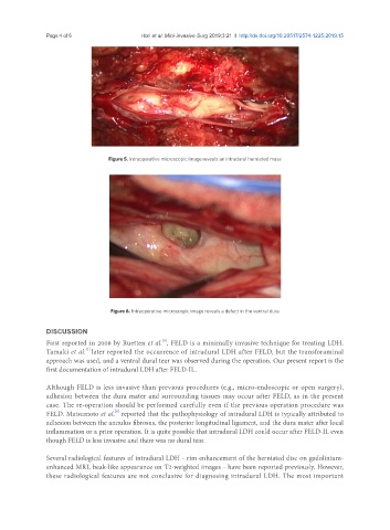
Figure 5. Intraoperative microscopic image reveals an intradural herniated mass
Figure 6. Intraoperative microscopic image reveals a defect in the ventral dura
DISCUSSION
[6]
First reported in 2008 by Ruetten et al. , FELD is a minimally invasive technique for treating LDH.
[7]
Tamaki et al. later reported the occurrence of intradural LDH after FELD, but the transforaminal
approach was used, and a ventral dural tear was observed during the operation. Our present report is the
first documentation of intradural LDH after FELD-IL.
Although FELD is less invasive than previous procedures (e.g., micro-endoscopic or open surgery),
adhesion between the dura mater and surrounding tissues may occur after FELD, as in the present
case. The re-operation should be performed carefully even if the previous operation procedure was
[8]
FELD. Matsumoto et al. reported that the pathophysiology of intradural LDH is typically attributed to
adhesion between the annulus fibrosus, the posterior longitudinal ligament, and the dura mater after local
inflammation or a prior operation. It is quite possible that intradural LDH could occur after FELD-IL even
though FELD is less invasive and there was no dural tear.
Several radiological features of intradural LDH - rim enhancement of the herniated disc on gadolinium-
enhanced MRI, beak-like appearance on T2-weighted images - have been reported previously. However,
these radiological features are not conclusive for diagnosing intradural LDH. The most important