
Page 178 - Read Online
P. 178
Page 2 of 5 Herbella et al. Mini-invasive Surg 2019;3:22 I http://dx.doi.org/10.20517/2574-1225.2019.19
Figure 1. Ports placement for operations on the esophagogastric junction. Liver retraction is moved from the right flank of the classic
approach to the epigastrium in the proposed didactic technique
A B
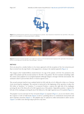
Figure 2. Esophageal exposition based on pre-taught hand positions of the second assistant. Exposure of the right side of the esophagus -
Position A, and exposure of the left side of the esophagus - Position B
METHOD
Ports are placed in a similar fashion to the classic approach with the exception of the liver retraction port
that is moved from the right flank to the epigastrium, closing to the xyphoid appendix [Figure 1].
The surgeon (chief resident/fellow) stands between the legs of the patient, with the first assistant to the
right of the patient and the second assistant to the left of the patient. The first assistant (attending) holds
the camera and a palpator (or an irrigator/aspirator although fatigue is stronger with this instrument). The
assistant is sited to prevent their elbows to touch surgeon’s arms.
The second assistant (2nd/3rd year resident) holds the EGJ with the aid of a Babcock or later on a Penrose
drain encircling the esophagus. The assistant is instructed at the beginning of the operation to place the
EGJ either in “position 1” or “position 2” as required. Position 1 exposes the right side of the esophagus
pointing the tip of the Babcock at the left inguinal area of the patient. Oppositely, position 2 exposes the
left side of the esophagus pointing the tip of the Babcock at the right inguinal area of the patient [Figure 2].
The palpator is active and used for liver retraction [Figure 3], exposure [Figure 4], as an extra hand to allow
the surgeon to work by both hands [Figure 5] and as a pointing device to communicate instructions and
show anatomic structures [Figure 6]. The palpator may be replaced by other instruments to allow suction
[Figure 5] or hold a knot during tying to prevent a slip knot.