
Page 173 - Read Online
P. 173
Hori et al. Mini-invasive Surg 2019;3:21 I http://dx.doi.org/10.20517/2574-1225.2019.15 Page 3 of 6
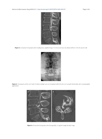
Figure 2. Computed tomography after myelography, sagittal image. An intradural mass was observed from L4 to the sacral level
Figure 3. Discography at the L4/5 level. Contrast medium was not contained within the disc as it spread intrathecally with a myelographic
appearance
A B
Figure 4. Computed tomography after discography. A: sagittal image; B: axial image