
Page 172 - Read Online
P. 172
Page 2 of 6 Hori et al. Mini-invasive Surg 2019;3:21 I http://dx.doi.org/10.20517/2574-1225.2019.15
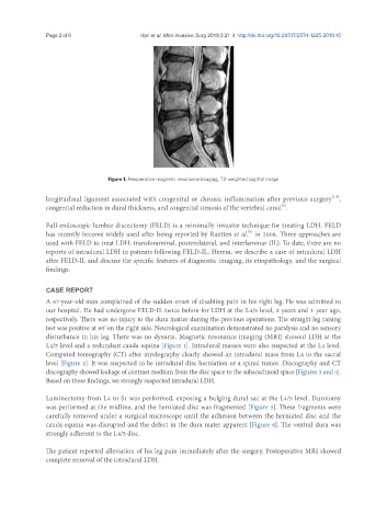
Figure 1. Preoperative magnetic resonance imaging, T2-weighted sagittal image
[1,4]
longitudinal ligament associated with congenital or chronic inflammation after previous surgery ,
congenital reduction in dural thickness, and congenital stenosis of the vertebral canal .
[5]
Full-endoscopic lumbar discectomy (FELD) is a minimally invasive technique for treating LDH. FELD
[6]
has recently become widely used after being reported by Ruetten et al. in 2008. Three approaches are
used with FELD to treat LDH: transforaminal, posterolateral, and interlaminar (IL). To date, there are no
reports of intradural LDH in patients following FELD-IL. Herein, we describe a case of intradural LDH
after FELD-IL and discuss the specific features of diagnostic imaging, its etiopathology, and the surgical
findings.
CASE REPORT
A 67-year-old man complained of the sudden onset of disabling pain in his right leg. He was admitted to
our hospital. He had undergone FELD-IL twice before for LDH at the L4/5 level, 2 years and 1 year ago,
respectively. There was no injury to the dura matter during the previous operations. The straight leg raising
test was positive at 60 on the right side. Neurological examination demonstrated no paralysis and no sensory
°
disturbance in his leg. There was no dysuria. Magnetic resonance imaging (MRI) showed LDH at the
L4/5 level and a redundant cauda equina [Figure 1]. Intradural masses were also suspected at the L4 level.
Computed tomography (CT) after myelography clearly showed an intradural mass from L4 to the sacral
level [Figure 2]. It was suspected to be intradural disc herniation or a spinal tumor. Discography and CT
discography showed leakage of contrast medium from the disc space to the subarachnoid space [Figures 3 and 4].
Based on these findings, we strongly suspected intradural LDH.
Laminectomy from L4 to S1 was performed, exposing a bulging dural sac at the L4/5 level. Durotomy
was performed at the midline, and the herniated disc was fragmented [Figure 5]. These fragments were
carefully removed under a surgical microscope until the adhesion between the herniated disc and the
cauda equina was disrupted and the defect in the dura mater apparent [Figure 6]. The ventral dura was
strongly adherent to the L4/5 disc.
The patient reported alleviation of his leg pain immediately after the surgery. Postoperative MRI showed
complete removal of the intradural LDH.