
Page 194 - Read Online
P. 194
Page 6 of 10 Zhao et al. Mini-invasive Surg 2018;2:26 I http://dx.doi.org/10.20517/2574-1225.2018.27
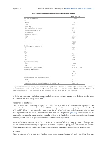
Table 1. Patient and lung tumour characteristics at repeat ablation
Total (n = 13)
Age (years), Mean (SD) 72 (11.0)
Sex
Female 7 (54)
Male 6 (46)
Tumour type
Non-small cell lung carcinoma 9 (69)
Colorectal adenocarcinoma 3 (23)
Pelvic sarcoma 1 (8)
Type of prior ablation/s
MWA 9 (69)
RFA 4 (31)
Number of prior ablation/s
1 11 (85)
2 2 (15)
Radiotherapy prior to ablations
No 9 (69)
Yes 4 (31)
Lobe
LLL 2 (15)
LUL 8 (62)
RLL 1 (8)
RUL 2 (15)
Repeat ablation modality
MWA 11 (85)
RFA 2 (15)
†
Diameter (mm) , median (IQR) 28.2 (22.0-40.0)
Number of ablation cycles
1 1 (8)
2 8 (62)
3 4 (31)
Maximum power used (Watts) (MWA, n = 11)
60-80 3 (27)
100-120 8 (73)
Total ablation duration (minutes) (MWA, n = 11), median (IQR) 4.5 (4.0-6.0)
† longest axial diameter in millimetres, immediately prior to repeat ablation. Data are presented as n (%) for categorical variables and
as mean [standard deviation (SD)] or median [interquartile range (IQR)] for continuous variables. MWA: microwave ablation; RFA:
radiofrequency ablation; LUL: left upper lobe; LLL: left lower lobe; RUL: right upper lobe; RLL: right lower lobe
of death was pulmonary embolism or myocardial infarction, however autopsy was declined and the cause
[11]
of death was not definitively determined .
Response to treatment
Only 12 patients had follow-up imaging performed. The 1 patient without follow-up imaging had died
2 days after the procedure. Median length of CT follow-up was 26 months (range 3-62), and median length
of FDG-PET follow-up was 6 months (range 0-26). Ten of twelve (83%) patients had complete ablation from
their repeat ablation procedure. Two of twelve (17%) had local progression [Table 2], one of whom had the
technically unsuccessful repeat ablation procedure. Time to first detection of local progression on imaging
for the 2 patients with local progression were 8 and 27 months respectively.
Six of twelve (50%) patients had nodal or distant metastasis on follow-up imaging. Four of these patients
had subsequent chemotherapy (the 2 patients in the local progression group, and 2 patients in the complete
ablation group). Median time to first detection of metastasis on imaging was 20 months (range 11-36).
Survival
Of all 13 patients, 8 (62%) were alive [median follow-up 30 months (range 3-91)] and 5 (38%) had died (me-