
Page 191 - Read Online
P. 191
Zhao et al. Mini-invasive Surg 2018;2:26 I http://dx.doi.org/10.20517/2574-1225.2018.27 Page 3 of 10
A B
C
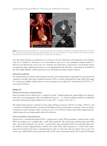
Figure 1. 57-year-old female with RUL SCC. A: Coronal FDG-PET scan 12 months post definitive radiotherapy (58 Gy) RUL T2N0 SCC;
B, C: microwave antenna (Acculis standard pMTA) centrally within the tumour; a total of 7 min 30 s ablation time-5 min 30 s at 80 W
followed by 2 min at 100 W. RUL: right upper lobe; SCC: squamous cell carcinoma
sion after initial ablation was defined as an increase in the size (maximum axial diameter) of the ablation
[9]
zone on CT. Stability or decrease in size of the ablation zone on CT was considered complete ablation .
Patients were followed up until 30 Jun 2017. Patients were either alive, evidenced by a documented visit to
an outpatient clinic, general practitioner or scan appointment after this date, or deceased, as recorded on
the state health database. Follow-up periods were calculated from date of repeat ablation.
Statistical methods
The characteristics of patients and treatment outcomes were summarised using frequencies and percent for
categorical variables and mean [standard deviation (SD)] or median [interquartile range (IQR) and range]
[10]
for continuous variables. Kaplan-Meier survival estimates were produced for overall survival. STATA 15
was used for analyses.
RESULTS
Patient and tumour characteristics
From December 2009 to March 2017, 13 patients (6 male, 7 female) underwent repeat ablation of a lung tu-
mour due to local progression after prior ablation(s) [Figures 1-5]. All prior ablations had been technically
successful. Mean age at repeat ablation was 72 years (SD 11, range 53-88) [Table 1].
The ablated lung tumours consisted of: non-small cell lung carcinoma (NSCLC) (8 stage 1 NSCLCs, and
1 multifocal bronchioloalveolar carcinoma with bilateral lung involvement), metastatic colorectal adeno-
carcinoma (3) and metastatic pelvic sarcoma (1). Four of the NSCLC lesions had locally progressed post-
radiotherapy and were subsequently referred for ablation.
Thermal ablation characteristics
Eleven of the 13 patients had repeat MWA; 7 patients after 1 prior MWA procedure, 2 patients after 2 prior
MWA procedures, and 2 patients after 1 prior RFA treatment. The remaining 2 patients had repeat RFA
after 1 prior RFA treatment. Nine of the 11 repeat MWAs were performed with the Acculis Microwave Tis-
sue Ablation system (Angiodynamics, Latham, New York, USA), which operates at 2.45 GHz with a maxi-
mum power output of 140 watts; a standard antenna with a 16 mm active tip was used in these 9 cases. The
remaining 2 repeat MWAs were performed with the HS Amica microwave system (HS Amica, HS Hospital