
Page 193 - Read Online
P. 193
Zhao et al. Mini-invasive Surg 2018;2:26 I http://dx.doi.org/10.20517/2574-1225.2018.27 Page 5 of 10
A C
B D
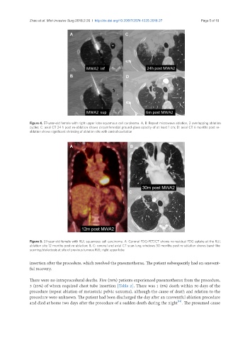
Figure 4. 57-year-old female with right upper lobe squamous cell carcinoma. A, B: Repeat microwave ablation, 2 overlapping ablation
cycles; C: axial CT 24 h post re-ablation shows circumferential ground-glass opacity of at least 1 cm; D: axial CT 6 months post re-
ablation shows significant shrinking of ablation site with central cavitation
A B
C
Figure 5. 57-year-old female with RUL squamous cell carcinoma. A: Coronal FDG-PET/CT shows no residual FDG uptake at the RUL
ablation site 12 months post re-ablation; B, C: coronal and axial CT scan lung windows 30 months post re-ablation shows band-like
scarring/atelectasis at site of previous tumour. RUL: right upper lobe
insertion after the procedure, which resolved the pneumothorax. The patient subsequently had an unevent-
ful recovery.
There were no intraprocedural deaths. Five (38%) patients experienced pneumothorax from the procedure,
3 (23%) of whom required chest tube insertion [Table 2]. There was 1 (8%) death within 30 days of the
procedure (repeat ablation of metastatic pelvic sarcoma), although the cause of death and relation to the
procedure were unknown. The patient had been discharged the day after an uneventful ablation procedure
[11]
and died at home two days after the procedure of a sudden death during the night . The presumed cause