
Page 89 - Read Online
P. 89
Chiu Modified parasternal approach for aortic valve surgery
time, blood product consumption and overall mortality were reduced. Conclusion: Reviewing the parasternal aortic valve series of
more than 500 cases, parasternal approach is safe, effective, and reproducible. The surgical trauma and blood product consumption were
minimized with this approach. Multiple valve procedures and ablation for atrial fibrillation are also feasible. Stable sternoclavicular
joints could facilitate early and aggressive activity of upper extremities for improved postoperative recovery. This approach could be a
good alternative option in aortic or multiple valve surgical procedures.
INTRODUCTION approach. Patients with isolated mitral procedures or
mitral and tricuspid procedures were done through
Minimally invasive cardiac surgery (MICS) has been right lateral mini-thoracotomy. For patients with aortic
widely adopted. Partial sternotomy for aortic valve valve involvement, Cosgrove’s idea was adopted
[7]
[1]
replacement is the most common MICS for aortic and modified with no rib resection. Initially, the two-rib
valve replacement. For multiple valve procedure, approach was initiated with the following major selection
[2]
full sternotomy still remains the choice for most criteria: adult single aortic valve cases, without chest
cardiac surgeons. There are several sternum-sparing wall deformity, without severe chronic obstructive
approaches, such as the anterior thoracotomy, [3,4] lateral pulmonary disease, and without aneurysmal or aortoiliac
thoracotomy and right parasternotomy. Among occlusive disease. Every single patient considered for
[6]
[5]
these three approaches, we modify the Cosgrove’s this approach had pre-operative chest, abdomen and
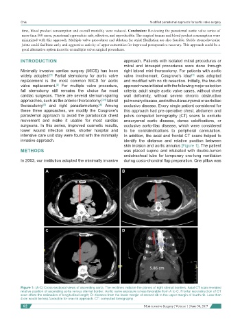
parasternal approach to avoid the paradoxical chest pelvis computed tomography (CT) scans to exclude
movement and make it usable for most cardiac aneurysmal aortic disease, dense calcifications, or
surgeons. In this series, improved cosmetic results, occlusive aorto-iliac disease, which were considered
lower wound infection rates, shorter hospital and to be contraindications to peripheral cannulation.
intensive care unit stay were found with the minimally In addition, the axial and frontal CT scans helped to
invasive approach. identify the distance and relative position between
skin incision and aortic annulus [Figure 1]. The patient
METHODS was placed supine and intubated with double-lumen
endotracheal tube for temporary one-lung ventilation
In 2003, our institution adopted the minimally invasive during costo-chondral flap preparation. One pillow was
A B
C D
Figure 1: (A-C) Cross-sectional views of ascending aorta. The red lines indicate the planes of right sternal borders. Axial CT scan revealed
relative position of ascending aorta versus sternal border. Aortic valve exposure is less favorable from A to C. Frontal reconstruction of CT
scan offers the estimation of longitudinal length; D: distance from the lower margin of second rib to the upper margin of fourth rib. Less than
4 cm would be less favorable for one-rib approach. CT: computed tomography
82 Mini-invasive Surgery ¦ Volume 1 ¦ June 30, 2017