
Page 162 - Read Online
P. 162
Bianchi et al. Modified fundoplication after Heller miotomy
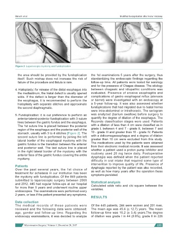
Figure 2: Laparoscopic myotomy and fundoplication
the area should be provided by the fundoplication the 1st examinations 5 years after the surgery, thus
itself. Such mishap does not increase the risk of standardizing the endoscopic findings regarding the
failure of the procedure and fistula is rare. follow-up time. All patients were tested for serology
and for the presence of Chagas disease. The etiology
4. Hiatoplasty: for release of the distal esophagus into between chagasic and idiopathic conditions was
the mediastinum, the hiatal defect is usually opened evaluated. Presence of erosive esophagitis and
wide. If the defect is larger than the diameter of complications of gastro esophageal reflux (stenosis
the esophagus, it is recommended to perform the or barret) were investigated with an endoscopy at
hiatoplasty with separate stitches and approximate a 5-year follow-up. It was also assessed whether
the second diaphragmatic. fundoplications that had migrated due to hiatal hernia
were intra-abdominal or intrathoracic. The seriogram
5. Fundoplication: it is our preference to perform an was analyzed (barium swallow) before surgery to
anterior-lateral-posterior fundoplication with 3 suture quantify the degree of dilation of the esophagus. The
lines between the gastric fundus and the esophagus. Rezende classification stages were used. Patients
The 1st suture line is placed between the posterior with a dilation of less than 4 cm were classified as in
region of the esophagus and the posterior wall of the grade I, between 4 and 7 - grade II, between 7 and
stomach, usually with 3 to 4 stitches [Figure 2]. The 10 - grade III and greater than 10 - grade IV. Patients
second suture line is performed by joining the left with a dolicomegaesophagus and a degree of dilation
lateral border of the esophageal myotomy with the greater than 10 cm were excluded from this study.
gastric fundus in the transition between the anterior The medications used by the patients were obtained
and posterior wall. The last suture line is placed from their electronic medical records. It was assessed
in the right lateral border of the myotomy with the whether a patient used a proton pump inhibitor and
anterior face of the gastric fundus covering the entire routinely used 20 mg twice daily. Postoperative
myotomy. dysphagia was defined when the patient reported
difficulty in oral intake that required some type of
Patients intervention to improve quality of life. Presence of
dysphagia reported by the patient was also recorded,
Over the past several years, the 1st choice of as well as how many years after the operation these
treatment for achalasia in our institution has been symptoms persisted.
the myotomy with fundoplication. Of the 849 patients
submitted to laparoscopic surgery between 2000 Statistical analysis
and 2012, 445 had regular follow-ups at our hospital Calculated odds ratio and chi square between the
for more than 5 years and underwent routine upper variables.
endoscopies. The examinations were performed every
2 years, or less if the patient presented any symptoms.
RESULTS
Data collection
The medical records of these patients were Of the 445 patients, 244 were women and 201 men.
reviewed and the following data were obtained: The mean age was 45.6 (± 12.7) years. The mean
age, gender and follow-up time. Regarding the follow-up time was 10.2 (± 3.4) years.The degree
endoscopy examinations, it was decided to analyze of dilation was grade I in 44 (9.8%), grade II in 226
Mini-invasive Surgery ¦ Volume 1 ¦ December 28, 2017 155