
Page 32 - Read Online
P. 32
Page 4 of 10 Mitura et al. Mini-invasive Surg 2021;5:22 https://dx.doi.org/10.20517/2574-1225.2021.19
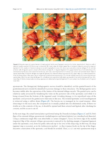
Figure 2. Desarda repair of a groin hernia in female patient (with round ligament resection for better visualization). Anterior wall of
inguinal canal is opened (A); hernia sac is dissected (B); lower edge of medial/upper flap of external oblique is sutured to inguinal
ligament (C, D); creation of external oblique strip with longitudinal incision (E); the strip is sutured to internal oblique (F, G); and the
anterior wall of inguinal canal is closed with external oblique (H, I). Inguinal ligament (1); upper/medial flap of external oblique (2);
undissected hernia sac [(3a) dissected hernia sac; and (3b) round ligamentum of uterus] (3); suture line between lower edge of
upper/medial flap of external oblique and inguinal ligament (4); external oblique aponeurosis (5); upper edge of a created aponeurotic
strip (6); lower edge of the upper flap of remaining incised external aponeurosis (7); internal oblique muscle (8); suture line between
upper edge of the strip and internal oblique (9); completed and sutured on both edges aponeurotic strip reinforcing inguinal floor (10);
lower flap of initially incised external oblique aponeurosis attached to inguinal ligament (11); and suture line of anterior wall of inguinal
canal (12).
aponeurosis. The ilioinguinal, iliohypogastric nerves should be identified, and the genital branch of the
genitofemoral nerve should be identified to prevent damage to these structures. The iliohypogastric nerve
becomes visible after the separation of the lamina of the internal oblique muscle. The genital nerve can be
relatively easily protected by visualizing the veins on the posterior side of the spermatic cord while it is
being released from the bottom of the inguinal canal. Avoiding damage to the superficial veins of the
spermatic cord protects the genital nerve that runs in this area. The isolated spermatic cord/round ligament
is retracted using a rubber drain [Figure 2B]. The hernia sac is managed in the usual manner. After
dissecting it to the neck area, the unopened sac is usually pushed into the abdominal cavity. If there are
doubts as to the contents of the sac, it should be opened and then sutured and ligated, after checking the
content, and the excess is cut off.
In the next stage, the actual hernioplasty is performed using the Desarda technique [Figure 2C and D]. Both
flaps of the external oblique aponeurosis (medial/superior and lateral/inferior) are visualized and dissected.
Using a continuous single-fiber non-absorbable 2.0 suture (Surgipro®, Tyco), the lower edge of the medial
(superior) flap of the external oblique aponeurosis is sutured to the shelving margin of inguinal ligament
under the spermatic cord, from the pubic tubercule up to the level of the deep ring. In this way, the deep
inguinal ring is recreated, as in the Lichtenstein method - so that it passes freely on the tip of the finger.
Excessive constriction of the spermatic cord should be avoided. Then, a 2-2.5 cm wide aponeurosis strip is