
Page 31 - Read Online
P. 31
Mitura et al. Mini-invasive Surg 2021;5:22 https://dx.doi.org/10.20517/2574-1225.2021.19 Page 3 of 10
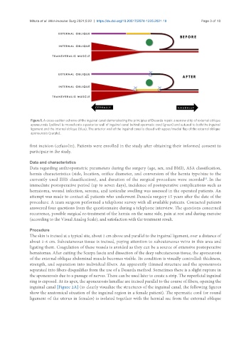
Figure 1. A cross-section scheme of the inguinal canal demonstrating the principles of Desarda repair: a narrow strip of external oblique
aponeurosis (yellow) is moved onto a posterior wall of inguinal canal behind spermatic cord (green) and sutured to both the inguinal
ligament and the internal oblique (blue). The anterior wall of the inguinal canal is closed with upper/medial flap of the external oblique
aponeurosis (purple).
first incision (cefazolin). Patients were enrolled in the study after obtaining their informed consent to
participate in the study.
Data and characteristics
Data regarding anthropometric parameters during the surgery (age, sex, and BMI), ASA classification,
hernia characteristics (side, location, orifice diameter, and conversion of the hernia type/size to the
currently used EHS classification), and duration of the surgical procedure were recorded . In the
[3]
immediate postoperative period (up to seven days), incidence of postoperative complications such as
hematoma, wound infection, seroma, and testicular swelling was assessed in the operated patients. An
attempt was made to contact all patients who underwent Desarda surgery 15 years after the date of the
procedure. A team surgeon performed a telephone survey with all available patients. Contacted patients
answered four questions from the questionnaire during a telephone interview. The questions concerned
recurrence, possible surgical re-treatment of the hernia on the same side, pain at rest and during exercise
(according to the Visual Analog Scale), and satisfaction with the treatment result.
Procedure
The skin is incised at a typical site, about 2 cm above and parallel to the inguinal ligament, over a distance of
about 5-6 cm. Subcutaneous tissue is incised, paying attention to subcutaneous veins in this area and
ligating them. Coagulation of these vessels is avoided as they can be a source of extensive postoperative
hematomas. After cutting the Scarpa fascia and dissection of the deep subcutaneous tissue, the aponeurosis
of the external oblique abdominal muscle becomes visible. Its condition is visually controlled: thickness,
strength, and separation into individual fibers. An apparently thinned structure and the aponeurosis
separated into fibers disqualifies from the use of a Desarda method. Sometimes there is a slight rupture in
the aponeurosis due to a passage of nerves. These can be used later to create a strip. The superficial inguinal
ring is exposed. At its apex, the aponeurosis lamellae are incised parallel to the course of fibers, opening the
inguinal canal [Figure 2A] (to clearly visualize the structures of the inguinal canal, the following figures
show the anatomical situation of the inguinal region in a female patient). The spermatic cord (or round
ligament of the uterus in females) is isolated together with the hernial sac from the external oblique