
Page 30 - Read Online
P. 30
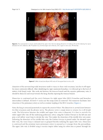
Page 6 of 13                            Gharagozloo et al. Mini-invasive Surg 2020;4:55  I  http://dx.doi.org/10.20517/2574-1225.2020.42


               Figure 5. The most posterior arm is used to retract the upper lobe inferiorly during dissection of stations 2R and 4R, clearing the space
               between the SVC anteriorly, the TR posteriorly, and the azygos vein inferiorly. SVC: superior vena cava; TR: trachea


                                   Figure 6. Further dissection of Level #10 node off the upper lobe bronchus (B)

               dissection of the surrounding nodal structures result in injury to these vital structures, but also it will repair
               the injury extremely difficult. After identifying the right mainstem bronchus, it is followed up to the level of
               station #10R lymph node. This node sits between the truncus branch and the superior pulmonary vein. It
               should be dissected and swept towards the lung, thereby exposing the truncus branch.


               Dissection is continued and the crotch between the right upper lobe (RUL) bronchus and bronchus
               intermedius is defined. All level #7 nodes and the sump node are removed. This maneuver facilitates later
               dissection of the pulmonary artery as well as eventual stapling of the RUL bronchus [Figure 6].


               Next, the lung is retracted posteriorly to expose the anterior hilum. The dissection is carried down between
               the hilar structures and the phrenic nerve. The phrenic nerve is swept down to remove the #10R lymph
               node. The bifurcation between the middle and upper lobe veins is dissected. It is best to encircle the
               entire upper lobe vein off the underlying pulmonary artery using the Cadiere Forceps in the left arm and
               pass a red rubber vessel loop to elevate the vein. This makes the dissection of the middle lobe vein easier.
               Following the dissection of the middle lobe vein, the Cadiere Forceps is passed under the elevated upper
               lobe vein, the vessel loop is released and re-grasped thereby isolating the upper lobe vein. Dissection
               is continued and the proximal main pulmonary artery is exposed as it emerges from the pericardium.
               The upper lobe vein is divided using a vascular stapler either using the robot arm or passed through the