
Page 34 - Read Online
P. 34
Page 10 of 13 Gharagozloo et al. Mini-invasive Surg 2020;4:55 I http://dx.doi.org/10.20517/2574-1225.2020.42
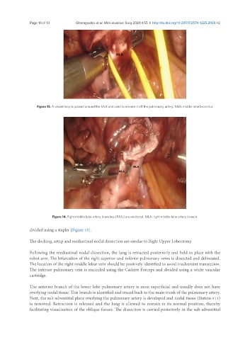
Figure 13. A vessel loop is passed around the MLB and used to elevate it off the pulmonary artery. MLB: middle lobe bronchus
Figure 14. Right middle lobe artery branches (RML) are encircled. MLA: right middle lobe artery branch.
divided using a stapler [Figure 15].
The docking, setup and mediastinal nodal dissection are similar to Right Upper Lobectomy.
Following the mediastinal nodal dissection, the lung is retracted posteriorly and held in place with the
robot arm. The bifurcation of the right superior and inferior pulmonary veins is dissected and delineated.
The location of the right middle lobar vein should be positively identified to avoid inadvertent transection.
The inferior pulmonary vein is encircled using the Cadiere Forceps and divided using a white vascular
cartridge.
The anterior branch of the lower lobe pulmonary artery is most superficial and usually does not have
overlying nodal tissue. This branch is identified and traced back to the main trunk of the pulmonary artery.
Next, the sub adventitial plane overlying the pulmonary artery is developed and nodal tissue (Station #11)
is removed. Retraction is released and the lung is allowed to remain in its normal position, thereby
facilitating visualization of the oblique fissure. The dissection is carried posteriorly in the sub adventitial