
Page 27 - Read Online
P. 27
Gharagozloo et al. Mini-invasive Surg 2020;4:55 I http://dx.doi.org/10.20517/2574-1225.2020.42 Page 3 of 13
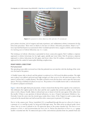
Figure 1. Port placement for robotic lobectomy in the right chest. AP: assistant port
poor patient selection, lack of surgeon and team experience, and rudimentary robotic instruments for the
[11]
lobectomy procedure. There were no deaths in the last 618 robotic lobectomy procedures. Meyer et al.
have reported that based on an assessment which included operative times, surgeon comfort, and mortality,
the learning curve of robotic lobectomy was 18 ± 3 cases.
The technique of robotic lobectomy is presented in three consecutive papers: Part I outlines a stepwise
approach to robotic lobectomy for the right upper, middle and lower lobes. Part II outlines a stepwise
approach to robotic lobectomy for left upper, and lower lobes. Part III outlines a methodical technical
approach for the control of catastrophic bleeding complications.
RIGHT-SIDED LOBECTOMY
Port placement
The operating room table is reversed such that the pedestal does not interfere with the docking of the robot
over the head of the patient.
A Double Lumen tube is placed, and the patient is positioned in a full lateral decubitus position. The right
arm is placed over pillows and positioned high enough such that access to the 4th intercostal space in the
anterior axillary line is readily attained. The table is flexed to move the hip down and to open the intercostal
spaces. The lung is deflated and placed on suction. The position of the double-lumen tube is rechecked after
the patient is prepped and draped.
Figure 1 shows the right chest port placement. A line is drawn from the tip of the scapula to the costal arch.
This delineates the highest point in the chest and the mid scapular line (posterior axillary line). Pleural
entry is with a Hassan Needle. Saline is infused and care is taken to look for easy egress of the saline from
the needle. If there is any question of pleural adhesions, we use a Visiport Instrument (Medtronic Inc.
Norwalk, Conn) for entry into the pleural space under direct vision. If the Visiport is used, a purse-string
is placed in the muscle layer and tied around the robot camera port to prevent CO leakage.
2
Port #1 is the camera port. Warm, humidified CO is insufflated through this port at a flow of 6 L/min to
2
a pressure of 6-8 mmHg to push the lung and diaphragm away. The other ports are placed under direct
vision. Port #2 (8 mm) is placed in the 7th intercostal space in the poster scapular line. This Port is 9 cm
posterior to Port #1 and accommodates da Vinci arm #2. Before the placement of Port #3, a 21-gauge
needle is inserted into the 7th intercostal space at the costovertebral junction from the patient’s back and