
Page 103 - Read Online
P. 103
Gharagozloo et al. Mini-invasive Surg 2021;5:39 https://dx.doi.org/10.20517/2574-1225.2021.74 Page 3 of 10
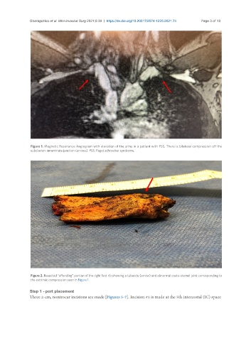
Figure 1. Magnetic Resonance Angiogram with elevation of the arms in a patient with PSS. There is bilateral compression off the
subclavian-innominate junction (arrows). PSS: Paget schroetter syndrome.
Figure 2. Resected “offending” portion of the right first rib showing a tubercle (arrow) and abnormal costo-sternal joint corresponding to
the extrinsic compression seen in Figure 1.
Step 1 - port placement
Three 2-cm, nontrocar incisions are made [Figures 5-7]. Incision #1 is made at the 5th intercostal (IC) space