
Page 105 - Read Online
P. 105
Gharagozloo et al. Mini-invasive Surg 2021;5:39 https://dx.doi.org/10.20517/2574-1225.2021.74 Page 5 of 10
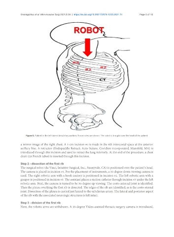
Figure 5. Patient in the left lateral decubitus position. Trocar sites are shown. The robot is brought over the head of the patient.
a mirror image of the right chest. A 1-cm incision #4 is made in the 6th intercostal space at the anterior
axillary line. A retractor (Endopaddle Retract; Auto Suture, Covidien incorporated, Mansfeld, MA) is
introduced through this incision and used to retract the lung inferiorly. At the end of the procedure, a chest
drain (28 French tubes) is inserted through this incision.
Step 2 - dissection of the first rib
The surgical robot (da Vinci, Intuitive Surgical, Inc., Sunnyvale, CA) is positioned over the patient’s head.
The camera is placed in incision #1. For the placement of instruments, a 30-degree down-viewing camera is
used. The right robotic arm with a hook cautery is positioned in incision #2. The left robotic arm with a
grasper is positioned in incision #3. The assistant places a suction catheter through incision #3 under the left
robotic arm. Next, the camera is turned to be 30-degree up viewing. The costo-asternal joint is identified.
Then the pleura overlying the first rib is dissected. The edges of the rib are identified, as is the costo-sternal
joint. Dissection of the pleura is carried just lateral to the subclavian artery. The lateral and posterior aspect
of the rib with the associated neurologic structures is left intact.
Step 3 - division of the first rib
Next, the robotic arms are withdrawn. A 30-degree Video-assisted thoracic surgery camera is introduced,