
Page 14 - Read Online
P. 14
Page 4 of 7 M’Harzi et al. Mini-invasive Surg 2019;3:27 I http://dx.doi.org/10.20517/2574-1225.2019.22
A B C
D E F
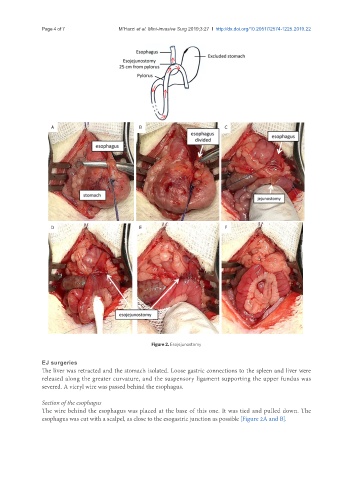
Figure 2. Esojejunostomy
EJ surgeries
The liver was retracted and the stomach isolated. Loose gastric connections to the spleen and liver were
released along the greater curvature, and the suspensory ligament supporting the upper fundus was
severed. A vicryl wire was passed behind the esophagus.
Section of the esophagus
The wire behind the esophagus was placed at the base of this one. It was tied and pulled down. The
esophagus was cut with a scalpel, as close to the esogastric junction as possible [Figure 2A and B].