
Page 61 - Read Online
P. 61
Page 4 of 13 Tang et al. Mini-invasive Surg 2024;8:24 https://dx.doi.org/10.20517/2574-1225.2024.04
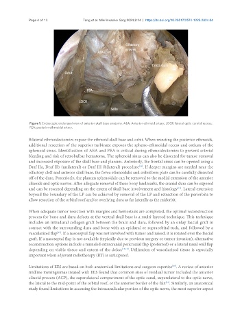
Figure 1. Endoscopic endonasal view of anterior skull base anatomy. AEA: Anterior ethmoid artery; LOCR: lateral optic carotid recess;
PEA: posterior ethmoidal artery.
Bilateral ethmoidectomies expose the ethmoid skull base and orbit. When resecting the posterior ethmoids,
additional resection of the superior turbinate exposes the spheno-ethmoidal recess and ostium of the
sphenoid sinus. Identification of AEA and PEA is critical during ethmoidectomies to prevent arterial
bleeding and risk of retrobulbar hematoma. The sphenoid sinus can also be dissected for tumor removal
and increased exposure of the skull base and planum. Anteriorly, the frontal sinus can be opened using a
[16]
Draf IIa, Draf IIb (unilateral) or Draf III (bilateral) procedure . If deeper margins are needed near the
olfactory cleft and anterior skull base, the fovea ethmoidalis and cribriform plate can be carefully dissected
off of the dura. Posteriorly, the planum sphenoidale can be removed to the medial extension of the anterior
clinoids and optic nerves. After adequate removal of these bony landmarks, the cranial dura can be exposed
and can be resected depending on the extent of skull base involvement and histology . Lateral extension
[14]
beyond the boundary of the LP can be achieved by removal of the LP and retraction of the periorbita to
allow resection of the orbital roof and/or overlying dura as far laterally as the midorbit.
When adequate tumor resection with margins and hemostasis are completed, the optimal reconstruction
process for bone and dura defects at the ventral skull base is a multi-layered technique. This technique
includes an intradural collagen graft between the brain and dura, followed by an onlay fascial graft in
contact with the surrounding dura and bone with an epidural or supraorbital tuck, and followed by a
vascularized flap . If a nasoseptal flap was not involved with tumor and raised, it is rotated over the fascial
[17]
graft. If a nasoseptal flap is not available (typically due to previous surgery or tumor invasion), alternative
reconstruction options include a tunneled extracranial pericranial flap (preferred) or a lateral nasal wall flap
depending on viable tissue and extent of the defect [18,19] . Utilization of vascularized tissue is especially
important when adjuvant radiotherapy (RT) is anticipated.
Limitations of EES are based on both anatomical limitations and surgeon expertise . A review of anterior
[20]
midline meningiomas treated with EES found that common sites of residual tumor included the anterior
clinoid process (ACP), the superolateral compartment of the optic canal, superolateral to the optic nerve,
the lateral to the mid-point of the orbital roof, or the anterior border of the falx . Similarly, an anatomical
[13]
study found limitations in accessing the intracanalicular portion of the optic nerve, the most superior aspect