
Page 60 - Read Online
P. 60
Tang et al. Mini-invasive Surg 2024;8:24 https://dx.doi.org/10.20517/2574-1225.2024.04 Page 3 of 13
bony lamina descends to form the perpendicular plate that articulates with the vomer and represents the
bony portion of the septum.
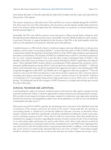
The anterior and posterior ethmoid arteries (AEA and PEA) are critical to identify during EES of SNMTs.
The AEA crosses the roof of the ethmoid bone after it travels across the superior-medial orbital wall at the
level of the posterior globe and approaches the cribriform plate in a posterior-to-anterior fashion from
lateral to medial [Figure 1].
Meanwhile, the PEA runs with the superior rectus and superior oblique muscle before exiting the orbit
through the posterior ethmoid canal and course horizontally across the cribriform plate in a more anterior-
to-posterior direction. A surgical landmark for the location of the PEA is the basal lamella, which lies
anterior to the attachment of the posterior ethmoid canal to the skull base.
Considerations prior to EES include a history of endonasal surgery, sinonasal inflammation or disease, prior
radiation, and the extent of neurological deficits . As previously discussed, for EES of SNMTs, additional
[5]
considerations include the histology and biological behavior of the SNMT, stage of disease, and anatomical
boundaries and limitations. The majority of SNMTs are epithelial tumors with SCC and adenocarcinoma as
[8]
the two most common histologies . SCC is also the most common histological type of SNMT in the
maxillary sinus while adenocarcinoma is the most common histology for SNMTs originating in the ethmoid
sinus . Other epithelial SNMTs include olfactory neuroblastoma (ONB), adenoid cystic carcinoma (ACC),
[9]
sinonasal undifferentiated carcinoma (SNUC), and sinonasal mucosal melanoma (SNMM) [10-12] . Different
tumors of the sinonasal cavity can also be classified by how aggressive the tumor is; low Hyams grade ONB
[10]
is typically less aggressive while SNUC and SNMM can be very aggressive with worse prognosis . Thus, the
decision to proceed with EES must depend on many factors and be compared to other treatment options
including open surgery and systemic treatment to ensure optimal outcomes for the patient. Induction
chemotherapy (IC) is increasingly being used for high-grade tumors such as SNUCs prior to definitive
therapy. The timing and overall role of surgery in SNMT treatment is a complex and evolving topic, beyond
the scope of this paper.
SURGICAL TECHNIQUE AND LIMITATIONS
Understanding the degree of exposure needed for gross total resection with negative margins and the
anatomical limitations of EES is critical for appropriate treatment selection and optimal patient outcomes.
EES provides several surgical technique advantages compared to traditional, open approaches including
limited brain retraction and manipulation, earlier tumor devascularization, and wide access to the skull base
including dura and optic nerves .
[13]
When performing EES for SNMTs, typically, the intranasal portion of the tumor is first debulked to provide
visualization of the margins and assess the extent of the tumor . Along with this, the opening of
[7]
surrounding sinuses is completed to provide the additional exposure necessary to determine tumor
margins, visualize the skull base, and identify critical bony landmarks including the carotid canals, optic
nerves, and lamina papyracea (LP) . When there is skull base involvement, often these landmarks are
[14]
included in the margins of resection, expanding the exposure from the posterior border of the frontal sinus
to the tuberculum sellae and between the medial wall of both orbits. When viable and not involved in
tumor, a nasoseptal flap is commonly raised from the contralateral side to the SNMT at the beginning of the
case and stored in either the maxillary sinus or nasopharynx until needed. In addition, a reverse rotational
flap can be raised to reconstruct the nasoseptal flap donor site if free of tumor . Nasal septal margins
[15]
should be checked to ensure that the reconstructive flap does not harbor residual microscopic tumor.